
Figures & data
Figure 1 Sequence analysis of the spike protein of camel MERS-CoV NRCE-HKU205. (A) Phylogenetic analysis of full-length spike protein of human and camel strains of MERS-CoV and related bat coronaviruses. The phylogenetic tree was generated using the Neighbor-Joining method (bootstrap resampling, 2000 replicates) with Geneious software from spike protein sequence alignment of representative MERS-CoV strains along with closely related bat betacoronaviruses retrieved from GenBank (accession numbers in methods section). Numbers near nodes represent percent consensus support. Scale bar represents estimated number of substitutions per site. camel, cam; human, hum. (B) Protein sequence alignment of coronavirus spike S2′ cleavage sites. Protein sequences of the S2′ cleavage site of representative coronaviruses from all four genera (accession numbers in methods section) were aligned using the ClustalW alignment method in Geneious software. Numbers indicate residue position within individual spike proteins.
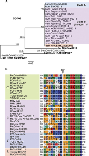
Figure 2 Effect of HKU205 S2′ substitutions on furin proteolytic cleavage. (A) Furin cleavage assay of fluorogenic peptides. Fluorogenic peptide mimetics of the S2′ spike cleavage sites of MERS-CoV strains EMC/2012 (blue line) and HKU205 (red line) were incubated with recombinant furin and the increase in fluorescence due to proteolytic processing was measured using a fluorometer. Assay performed in triplicates with results representing averages from three independent experiments of relative fluorescence units over time (n=3). Error bars indicate SD. (B) Analysis of the consequences of HKU205 S2′ substitutions on furin proteolytic processing products of MERS-CoV S by western blot. Murine leukemia virus (MLV) pseudovirions bearing EMC/2012 S protein with either a wild-type (EMCwt) or with a HKU205 substituted S2′ cleavage site (EMC205-S2′) were either untreated or treated with exogenous furin. The samples were analyzed by western blot to compare proteolytic cleavage of S proteins. The MLV capsid protein p30 was used as loading control.
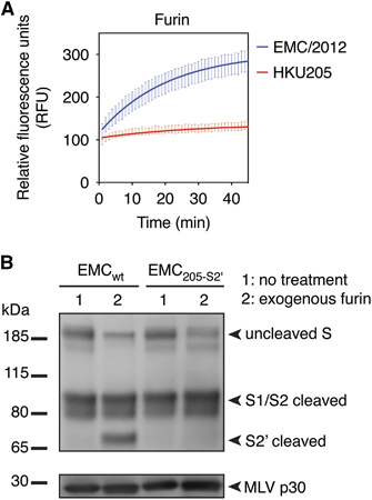
Table 1 Comparison of predicted and measured furin-mediated proteolytic processing of EMC/2012 and HKU205 MERS-CoV S2′ sites
Figure 3 Impact of HKU205 S2′ substitutions on fusion-activation and host cell entry mediated by MERS-CoV S. (A) Assessment of the effect of HKU205 S2′ substitutions on MERS-CoV S fusogenicity. Huh-7 cells were transfected to express MERS-CoV EMC/2012 S with either wild-type (EMCwt) or HKU205 substituted S2′ site (EMC205-S2′). The cells were either untreated, co-transfected with a furin-encoding plasmid (furin overexpression), treated with exogenous furin (furin exogenous) or treated with 75 μM of the furin inhibitor dec-RVKR-CMK (furin inhibitor). The cells were then processed for immunofluorescence with labeling of S protein (false colored green) and nuclei (DAPI, blue). Scale bar represents 100 μm. Quantitative microscopy analysis was undertaken to assess the average sizes of syncytia induced by S expression. Ten syncytia were analyzed for each condition and data are the averages of nuclei/syncytium from three independent experiments (n=3). (B) Effect of HKU205 S2′ substitutions on MERS-CoV S-mediated host cell entry. MLV-pseudotyped viruses harboring EMC/2012 S protein with either a wild-type (EMCwt) or with a HKU205 substituted S2′ cleavage site (EMC205-S2′) and containing a luciferase reporter gene were used to infect human Huh-7 (liver) and MRC-5 (lung) cells along with simian Vero-E6 (kidney) cells. 72 h post infection, cells were assayed for luciferase activity using a luminometer. Assays performed in triplicates and results are averages of relative luciferase units from two independent experiments (n=2). For both A and B, error bars indicate SD and statistical significance analyses were performed using two-tailed Student’s t-test. Not significant (NS), P>0.05; very highly significant, ***P≤0.001.
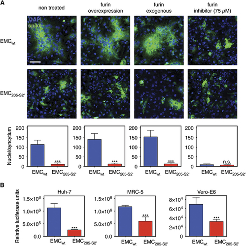
Figure 4 Analysis of proteolytic cleavage of HKU205 S2′ site by trypsin and cathepsin L. (A) Trypsin cleavage assay of fluorogenic peptides. Fluorogenic peptide mimetics of the S2′ spike cleavage sites of MERS-CoV strains EMC/2012 (blue line) and HKU205 (red line) were incubated with TPCK-treated trypsin and the increase in fluorescence due to proteolytic processing was measured using a fluorometer. (B) Cathepsin L cleavage assay of fluorogenic peptides. Assay performed as in A with cathepsin L used instead of trypsin. Both trypsin and cathepsin L cleavage assays were performed in triplicates with results representing averages of relative fluorescence units over time from three independent experiments (n=3). Error bars indicate SD.
Table 2 Summary of rates of proteolytic cleavage (V max) of EMC/2012 and HKU205 MERS-CoV S2′ sites
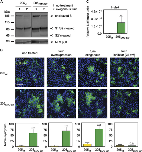
Figure 5 Effect of introducing the EMC/2012 S2′ furin cleavage site in HKU205 S. (A) Analysis of furin cleavage products. MLV-pseudotyped viruses bearing HKU205 S protein with either a wild type (205wt) or with a EMC/2012 substituted S2′ cleavage site (205EMC-S2′) were either untreated or treated with exogenous furin. The samples were analyzed by western blot to compare proteolytic cleavage products of S proteins. The MLV capsid protein p30 was used as loading control. (B) Impact of the introduction of the EMC/2012 S2′ furin cleavage site on HKU205 S-mediated fusion. Huh-7 cells were transfected to express MERS-CoV HKU205 S with either wild type (205wt) or EMC/2012 substituted S2′ site (205EMC-S2′). The cells were either untreated, co-transfected with a furin-encoding plasmid (furin overexpression), treated with exogenous furin (furin exogenous), or treated with 75 μM of the furin inhibitor dec-RVKR-CMK (furin inhibitor). The cells were then processed for immunofluorescence with labeling of S protein (false colored green) and nuclei (DAPI, blue). Scale bar represents 100 μm. Quantitative microscopy analysis was undertaken to assess the average sizes of syncytia induced by S expression. Ten syncytia were analyzed for each condition and data are the averages of nuclei/syncytium from three independent experiments (n=3). (C) Effect of introducing the EMC/2012 S2′ furin cleavage site on HKU205 S-mediated host cell entry. MLV-pseudotyped viruses harboring HKU205 S protein with either a wild type (205wt) or with a EMC/2012 S2′ furin cleavage site (205EMC-S2′) and containing a luciferase reporter gene were used to infect human Huh-7 (liver) cells. Seventy-two hours post infection, cells were assayed for luciferase activity using a luminometer. Assays performed in triplicates and results are averages of relative luciferase units from two independent experiments (n=2). For both B and C, error bars indicate SD and statistical significance analyses were performed using two-tailed Student's t-test. Not significant (NS), P>0.05; highly significant, **P≤0.01; very highly significant, ***P≤0.001.