Figures & data
Table 1 Mice and hamster study design
Figure 1 Immunogenicity and adjuvanticity of recombinant C. difficile flagellar protein FliC in mice. (A) 2 µg of purified recombinant C. difficile flagellar protein FliC was analyzed by SDS-PAGE on a gel stained with Coomassie blue (left panel). Whole cell lysate from uninduced and IPTG-induced BL21 (DE3)* cells were analyzed by Western blot using anti-His antibody (right panel). M, molecular weight markers; I, IPTG-induced cell lysate; U, uninduced cell lysate. Serum anti-FliC IgG (B), anti-TcdA IgG (C), and anti-TcdB IgG (D) responses in mice immunized on days 0, 14, and 28 in serum collected on days 14, 28, and 42. Cohorts of mice received 25 µg total of FliC adjuvanted with alum, unadjuvanted TcdARBD, and TcdBRBD or TcdARBD and TcdBRBD adjuvanted with FliC. Results were determined by kinetic ELISA and are reported as OD/min; the geometric mean plus standard error of the mean for each cohort is shown. * denotes statistical significance (P < 0.05), using an unpaired Student t-test analysis for comparison of means; for nonparametric data, we used the Mann–Whitney U-test.
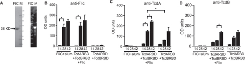
Figure 2 Survival and spore shedding in FliC-immunized mice following C. difficile UK1 challenge. (A) Survival in vaccinated C57BL/6 mice following orogastric challenge with 106 CFU of C. difficile strain UK1. Cohorts of mice received 25 µg total of FliC adjuvanted with alum, unadjuvanted TcdARBD, and TcdBRBD or TcdARBD and TcdBRBD adjuvanted with FliC. Control mice were immunized with saline. Mice were immunized on day 0, 14, and 28 and challenged two weeks after the last immunization following antibiotic treatment. Mantel–Cox test for pairwise comparisons and Gehan Breslow Wilcoxon test was used to assess statistical significance between the cohorts. (B) C. difficile shedding in mouse fecal samples from immunized and control mice following orogastric challenge with 106 CFU of C. difficile strain UK1. Results denote fecal shedding of C. difficile reported as CFU per gram (the geometric mean plus standard error of the mean) from fecal pellets. One fecal pellet was collected on days 3, 4, and 5 post-challenge from every mouse in each cohort containing five mice. * denotes statistical significance (P < 0.05), using an unpaired Student t-test analysis for comparison of means.
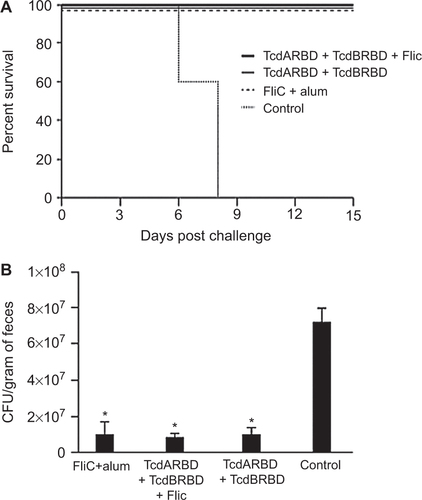
Figure 3 Anti-FliC IgG dose response and survival in FliC immunized mice challenged with C. difficile UK1. (A) Survival in vaccinated C57BL/6 mice following orogastric challenge with 106 CFU of C. difficile UK1. Cohorts of mice received either 5 µg or 25 µg total of FliC adjuvanted with alum. Control mice were immunized with saline. Mice were immunized on day 0, 14, and 28 (three immunizations) or on days 0 and 14 (two immunizations) and challenged two weeks after the last immunization following antibiotic treatment. Mantel–Cox test for pairwise comparisons and Gehan Breslow Wilcoxon test was used to assess statistical significance between the cohorts. (B) Percent weight change of surviving mice compared to prechallenge (baseline weight), day 2, day 4, and day 7 following orogastric challenge with 106 CFU C. difficile UK1. Results are reported as the geometric mean plus standard error of the mean for each cohort for each day. All mice in all cohorts are alive at day 2, all control mice succumb to CDI at day 7. * denotes statistical significance (P < 0.05), using an unpaired Student t-test analysis for comparison of means; when compared to the control cohort for each time point. (C) Anti-FliC IgG responses in serum of mice one day before challenge. Results were determined by kinetic ELISA and are reported as OD per minute; the geometric mean plus standard error of the mean for each cohort is shown. Data points circled denote mice that succumbed to challenge.) * denotes statistical significance (P < 0.05), using an unpaired Student t-test analysis for comparison of means; for nonparametric data, we used the Mann–Whitney U-test.
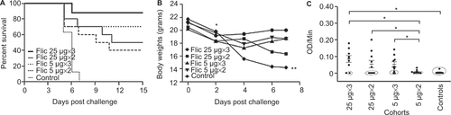
Figure 4 Protection of passively immunized C57BL/6 mice from challenge with C. difficile UK1. (A) Survival in passively immunized C57BL/6 mice following orogastric challenge with 106 CFU of C. difficile UK1. Cohorts of mice received either 400 µl total of hyperimmune FliC serum or naïve serum. Antibiotic-treated mice were transfused on day 0 and challenged 24 h later. Mantel–Cox test for pairwise comparisons and Gehan Breslow Wilcoxon test was used to assess statistical significance between the cohorts. (B) Detection of circulating anti-FliC IgG on days 1, 3, 7, and 10 in serum of mice passively immunized with hyperimmune FliC serum. Results were determined by kinetic ELISA and are reported as OD per minute. Numbers denote individual mice. Mouse 178 succumbed to challenge on day 7, therefore no serum sample was collected for days 7 and 10.
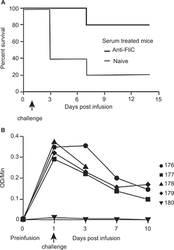
Figure 5 Experimental design of immunization and protective efficacy experiments in male Golden Syrian hamsters.
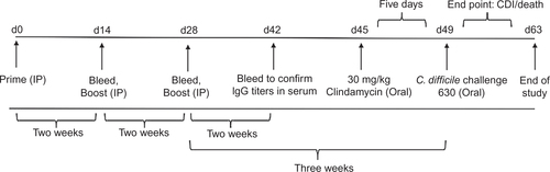
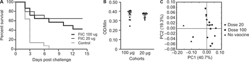
Figure 6 Survival in FliC-immunized golden Syrian hamsters following C. difficile 630Δerm challenge. (A) Survival in control and vaccinated golden Syrian hamsters following orogastric challenge with 500 CFU of C. difficile 630Δerm. Cohorts of hamsters received either 20 µg or 100 µg of FliC adjuvanted with alum. Control hamsters were immunized with saline. Mantel-Cox test for pairwise comparisons and Gehan Breslow Wilcoxon test was used to assess statistical significance between the cohorts. (B) Anti-FliC IgG responses in serum of vaccinated hamsters 1 week before challenge. Results were determined by kinetic ELISA and are reported as OD per minute; the geometric mean plus standard error of the mean for each cohort is shown. An unpaired Student t-test analysis was used for comparison of means. (C) PCoA of beta diversity present in the fecal bacterial communities in hamsters one week before challenge as measured by 16S rRNA. Animals that received no vaccine are represented by black triangles, 20 µg of vaccine are represented by circles, and 100 µg of vaccine are represented by squares.