Figures & data
Figure 1 Map depicting the geographical distribution of the BoDV-1-positive horses and shrews. The inset shows the area in Upper Austria, which has been enlarged.
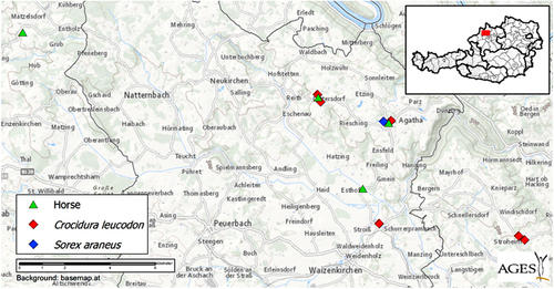
Table 1 Overview of clinical signs and histopathological findings in the affected horses
Table 2 Sequences of 13 primer pairs used in addition to previously described primers2 for the generation of complete BoDV-1 genomes
Figure 2 Bornavirus encephalitis in horses. (A) Severe nonpurulent encephalitis with perivascular cuffing (asterisk) and microgliosis. Bar=250 μm. (B and C) Eosinophilic intranuclear inclusion bodies (arrows) in a brainstem neuron (B) and a trigeminal ganglion neuron surrounded by lymphohistiocytic infiltration (C). Bars=25 μm. (D and E) Immunohistochemical demonstration of BoDV antigen (brown signals) in neurons and glial cells and their processes. Bars=100 μm. (A–C) Hematoxylin and Eosin staining. (D and E) Bo-18 immunohistochemistry.
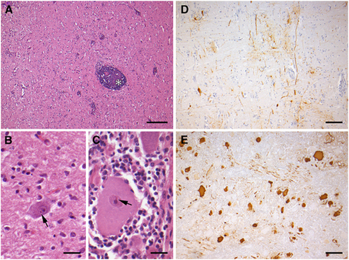
Figure 3 Bornavirus infection of shrews. (A and B) Demonstration of BoDV-1 antigen in an infected Crocidura leucodon shrew. In the brain (A), the antigen is diffusely distributed; in skin (B), keratinocytes and periadnexal nerve fibers are positive. (C and D) Demonstration of BoDV-1 antigen in an infected Sorex araneus shrew. In the brain (C), the viral antigen has a multifocal, patchy staining pattern, representing individually stained neurons and glial cells and their processes. In skin (D), the viral antigen is confined to periadnexal nerve fibers; keratinocytes are negative. (E and F) Lack of immunoreactivity in brain (E) and skin (F) in a non-infected C. leucodon shrew. Bars=40 μm; Bo-18 immunohistochemistry.
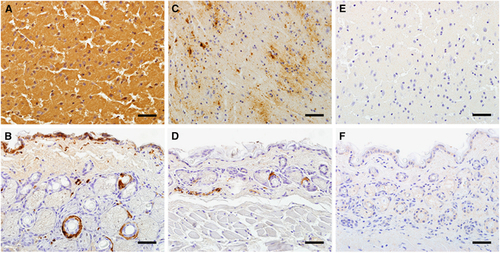
Figure 4 Phylogenetic tree of the complete coding sequences of representative members of the genus Bornavirus including new Upper Austrian sequences. (A) For better visualization, the eleven sequences determined in this study are collapsed. (B) Only the sequences of the new Upper Austrian Bornaviruses are presented. These sequences are marked with green triangles (horse-derived BoDVs-1), red diamonds (C. leucodon-derived BoDVs-1), and blue diamond (S. araneus-derived BoDVs-1). GenBank accession numbers, strain names (in the case of BoDVs) and species abbreviationsCitation3 are indicated at the branches. All supporting bootstrap values are displayed next to the nodes. The horizontal scale bar indicates genetic distances. *GenBank acc. nos: KY002971-79 and KY490040-41.
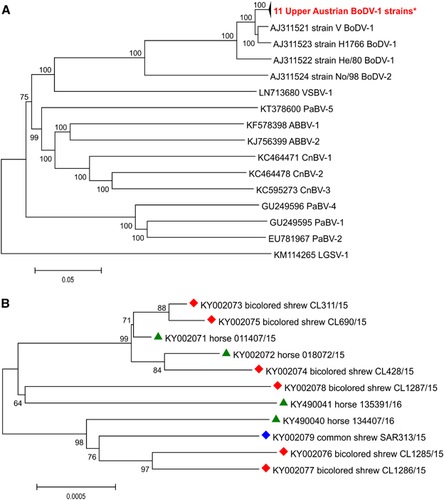
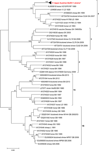
Figure 5 Phylogenetic tree of an 1824-bp long sequence stretch (coding for N protein, intergenic N/X, as well as for X and P proteins) of 43 selected BoDVs-1 from the GenBank and eleven sequences determined in this study (which are collapsed). The five major clusters are indicated. The GenBank accession numbers, host or strain names, geographic location and years of isolations are indicated at the branches. Supporting bootstrap values ≥70% are displayed next to the nodes. The horizontal scale bar indicates genetic distances (here 0.5% nucleotide sequence divergence). BA, Bavaria; BW, Baden-Wurttemberg; GB, Graubuenden; HE, Hesse; L, Liechtenstein; RP, Rhineland-Palatinate; SA, Saxony-Anhalt; SG, Sankt Gallen; SX, Saxony; TH, Thuringia. *GenBank acc. nos: KY002971-79 and KY490040-41.