Figures & data
Figure 1 Characterisation of FPV mutants. (A) Cleavage site motifs of WT and mutant viruses. The WT cleavage site sequence of FPV and the two mutants produced for the generation of recombinant viruses are depicted. Asterisks indicate the site of cleavage. (B and C) Replication of viruses in MDCK cell culture in the absence and presence of trypsin. (B) Cells were inoculated at a MOI of 0.001. At the indicated time points, culture supernatants were monitored for HA titres using chicken red blood cells. (C) Spread of virus infection traced by plaque formation. Monolayers seeded in six-well plates (diameter per well: 35 mm) were inoculated with the indicated viruses and covered with an agarose-containing medium overlay. At 3 days post-infection, plaques were visualized by staining cell monolayers with a crystal violet solution.
Figure 2 Analysis of cleavage activation of HA. MDCK cells were infected at a MOI of 5 and incubated in 35S-methionine/cysteine containing medium in the absence or presence of trypsin. After 12 h, metabolically labelled progeny viruses were collected from the cell supernatants by ultracentrifugation and aliquots of the virus preparations were applied to a 10% sodium dodecyl sulfate–polyacrylamide gel electrophoresis. Protein bands were visualized by fluorography. The uncleaved HA precursor (HA0) and the cleavage products HA1 and HA2 are indicated.
Figure 3 Pathogenicity and virus yields in embryonated chicken eggs. (A) Virus yields in allantoic fluids 30 h post-inoculation. (B) Mortality of embryos over time. Eleven-day-old embryonated eggs were inoculated by the allantoic route with 2000 pfu/egg. At the indicated time points post-inoculation, eggs were opened and checked visually for viability of embryos. The survival rates are given as per cent values based on the total number of eggs infected per virus species.
Figure 4 Spread of virus infection to embryonic tissues. Eleven-day-old embryonated chicken eggs were inoculated as outlined in the legend to . At 48 h post-inoculation, embryos were removed for the preparation of sagittal cryosections. Virus infection of the depicted tissues was detected by means of in situ hybridisation employing a digoxygenin-labelled riboprobe specific for the FPV-NP gene. Embryonic tissues were visualized by common haematoxylin–eosin staining (magnification: ×63).
Figure 5 Pathogenicity of FPV mutants in adult chicken. Three groups of ten 6-week old SPF chickens each were inoculated intravenously with fresh infective allantoic fluid (HA titre >24) containing WT, Mut1 or Mut2 viruses. From then on, birds were examined at 24-h intervals for the following 10 days. At each observation, the birds are examined for clinical signs of disease and scored 0 if normal, 1 if mildly sick, 2 if severely sick and 3 if dead (for details, see the section on ‘Materials and methods’). The intravenous PI is the mean score of birds per group over the entire observation period. PI, pathogenicity index.
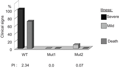
Table 1 Immunogenicity and protective efficacy in chickens of the inactivated vaccine prepared from FPV mutant 1
Figure 6 Immunisation of mice with formalin-inactivated vaccine prepared from FPV mutant 1 (H7N1) and subsequent challenge with 100LD50 of SC35M (H7N7). Groups of five mice each were immunized intramuscularly as indicated in the boxes. An unvaccinated group was used as a control. Mice were monitored for 14 days after immunisation (A) and after challenge (B) for weight loss and signs of disease.
