
Figures & data
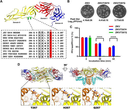
a Upper panel: crystal structure of the JEV E monomer with the αB helix highlighted in green (PDB: 3P54); lower panel: sequence alignment of the αB helices from the E proteins of different flaviviruses. The E-264 residue (numbering based on JEV) is highlighted in red. The other three conserved residues adjacent to E-264 are shown in gray. b Plaque morphologies and peak titers (N = 2) of ZIKV and the T267 mutants. c Thermostability analysis of ZIKV and the T267 mutants at 50 °C. ****P < 0.0001. N = 2. A one-way analysis of variance (ANOVA) was performed to analyze the significant differences between each treatment group and the corresponding untreated group, and the data were expressed as the mean ± standard deviation. d A potential interaction network centering on T267/H267/Q267 near helix αB was analyzed using the SWISS-MODEL Workspace in the Expasy web server. Top: side view of the atomic model of the E:M:M:E heterotetramer of ZIKV (PDB code: 5IRE) shown in ribbon. Domains I, II, III, and the transmembrane (TM) of E and M are shown in red, yellow, blue, cyan, and orange, respectively. Bottom: the detailed interaction networks centering on residue 267 from structures of the WT ZIKV and the T267 mutants. Structural figures were prepared with PyMol and UCSF Chimera version 1.10.1 (the Regents of the University of California)