Figures & data
List of H3N2 viruses isolated from canines in South Korea from 2009 to 2013
Amino acid sequence comparison between virus isolates
Differences in the amino acid residues of each gene are indicated by single-letter amino acid codes with positions indicated at the top of the diagram. The black and white letters indicate the amino acid regions originating from the first Korean CIV and recent CIVs, respectively. The amino acid positions of HA are based on the numbering of the H3 HA. Type I includes only one A/canine/Korea/AS-01/2009 virus. Three individual isolates (AS-05/12, AS-08/12, and AS-09/12 viruses) comprise Type II viruses. Type III viruses were the most divergent viruses from the Type I virus, containing six individual isolates (AS-03/12, AS-04/12, AS-06/12, AS-11/13, AS-14/13, and AS-15/13 viruses)
The body temperatures of infected (a) and contact (b) beagles (†succumbed to death) were monitored. Groups of four beagles inoculated with 105.5 EID50 of each virus were individually placed adjacent to naïve (contact) beagles. The mean viral titers (log10 EID50/mL) for nasal-wash (c) or lung and tracheal (d) samples are shown for each group of beagles. The limit of virus detection was 0.7 log10 EID50/mL or gram. Statistically significant differences (*P < 0.05, **P < 0.001, and ***P < 0.0001) between each representative canine and AS-01/09 viruses are indicated by *P value was determined using one-way ANOVA
Each virus was infected at an MOI of 0.001 in MDCK cells (a) or 0.01 in NHBE cells (b) in the presence of L-1-tosylamido-2-phenylethyl chloromethyl ketone (TPCK)-treated trypsin. Supernatants of virus cultured cell lines were harvested at 12, 24, 48, and 72 hpi, and virus titers were measured in MDCK cells. The virus titers were determined as means ± SD from three independently performed experiments. Dotted lines indicate the limit of detection (1.8log10TCID50/mL). Statistically significant differences (*P < 0.05, **P < 0.001, and ***P < 0.0001) between each representative canine and AS-01/09 viruses are indicated by *P value was determined using one-way ANOVA
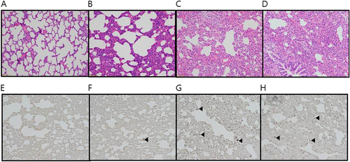
Ferrets were intranasally inoculated with 107.0 EID50/mL of each virus. Lungs were harvested 5 days after virus inoculation. LPM91/06 (a and e), AS-01/09 (b and f), AS-05/12 (c and g) and AS-11/13 (d and h), respectively. No damage was evident in lung sections from the LPM91/06-infected group (a and e). Moderate to severe lung damage, indicated by lesions, are shown in the AS-01/09 (b and f), AS-05/12 (c and g) and AS-11/13 (d and h) groups. Arrows indicate positive immunostaining
List of canine H3N2 RG viruses in the backbone of the LPM91/06 virus
Groups of four dogs were inoculated with 105.5 TCID50/mL of each reassorted virus (Table ). RD-contacts were individually placed adjacent to the inoculated dogs at 1 dpi. Nasal wash titers are shown for individual dogs (a and b). Two dogs per group were humanely euthanized at 5 dpi for virus titration in various tissues (c). Virus titers in nasal washes and homogenized tissues are expressed as log10EID50/mL or gram of tissue collected, with the limit of virus detection set at 0.7 log10 EID50/mL or gram