
Figures & data
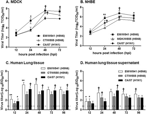
Replication of two Em/W541(H5N6) and CT/W555(H5N8) viruses were monitored in Madin-Darby Canine Kidney (MDCK) cells (a), Normal Human Bronchial Epithelial (NHBE) cells (b), Human lung tissue explants and corresponding culture supernatants (c and d, respectively) starting at 12 and 24 hpi intervals thereafter. Growth kinetics were compared to those of the control 2009 pandemic CA/07(H1N1) virus. The titers shown are means ± SD from three independently performed experiments. (*p < 0.05, **p < 0.01, †p < 0.001)
Biological properties of HPAI H5 viruses
Virus replication was examined in chickens and ducks that had been experimentally inoculated via the intranasal route with 106 EID50/ml of each virus. Oropharyngeal swabs, cloacal swab, and lung titers of chickens are shown for Em/W541(H5N6) (a) and CT/W555(H5N8) (b). Oropharyngeal swabs, cloacal swabs, and lung titers from ducks are shown for Em/W541(H5N6) (c) and CT/W555(H5N8) (d). Mean viral titers (log10 EID50/ml) are shown for each group of birds. The limit of virus detection was 0.7 log10 EID50/ml
Viral tissue titers in experimentally inoculated animals
Concentrations of various cytokines/chemokines in BAL fluid from mice at 1, 3, 5, and 7 dpi were measured by protein analysis with the Luminex™ Instrumentation Systems multiplex array reader (Bio-Plex Workstation from Bio-Rad Laboratories). The values shown are means ± SD (error bars) from BAL fluid of three mice per time point tested. (*p < 0.05)
Fig. 4 Histopathology and immunohistochemical staining for influenza virus antigen in mouse (a, b, e, and f) and ferret (c, d, g, and h) lungs at 3 (a, c, e, and g) and 5 dpi (b, d, f, and h) with HPAI Em/W541(H5N6) and CT/W555(H5N8).In the lung, viral antigens are widely presented in the alveolar septum which are mildly thickened with increased cellularity of inflammatory cells and also necrotic cell debris are presented in the lumen (arrows in a, b, e, and f)
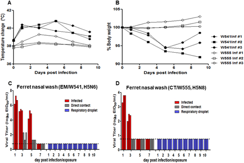
Rectal temperatures and body weights were measured at 1, 3, 5, 7, 9, 11, and 14 dpi (a and b). Individual nasal wash titers are shown for ferrets inoculated with Em/W541(H5N6) and CT/W555(H5N8). To examine transmission, the inoculated animals were individually paired with a direct contact (DC) and a respiratory droplet (RD)-contact animal (1:1:1 setup, triplicate) (c and d). The mean viral titers (log10 EID50/ml) are shown for each group of mammals. The limit of virus detection was 0.7 log10 EID50/ml
Seroconversion in inoculated and contact ferrets