Figures & data
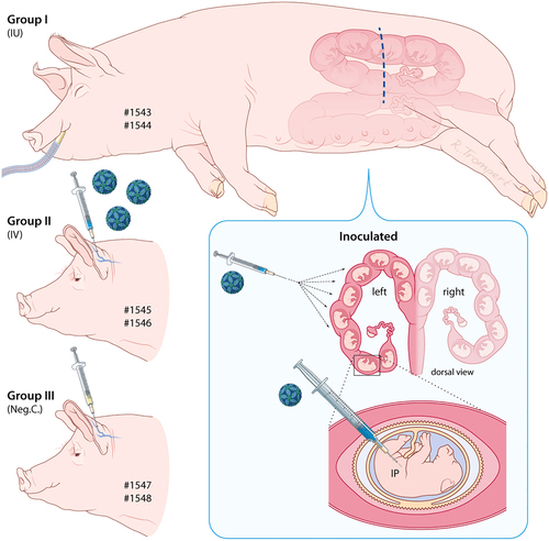
Sows 1543 and 1544 were subjected to laparotomy, after which all fetuses in the left uterine horn were inoculated, via IP route, with 105.6 TCID50 of ZIKV strain PRVABC59. Sows 1545 and 1546 were inoculated via IV route with 106.4 TCID50 of the same ZIKV strain and sows 1547 and 1548 were injected with culture medium, also via IV route
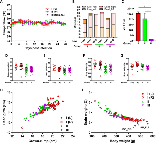
a Averages and standard deviations (SDs) of daily recorded sow body temperatures. b Numbers of live and dead (mummified) fetuses per sow and per uterine horn (left or right) at the time of necropsy. c ZIKV-specific VNT titers in sow serum at four weeks post challenge. Averages and SDs are presented. The limit of detection was set to 40. d–g Averages and SDs of crown-rump lengths d, head girths e, total weights f, and brain weights g of the live fetuses per experimental (sub)group. h, i Relative head size h and relative brain weight i of all individual fetuses. L left uterine horn,R right uterine horn. An asterisk indicates a statistical difference
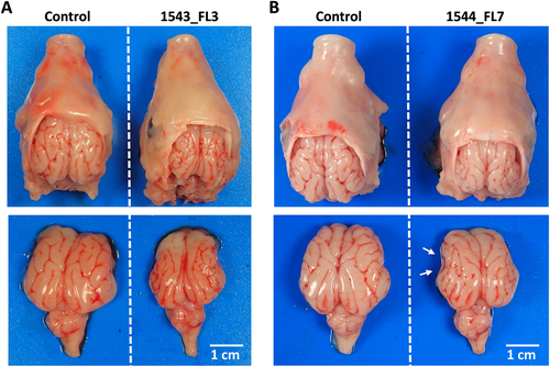
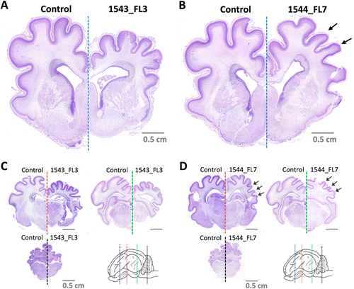
Top view of heads and brains of fetuses presenting stunted brain growth; 1543_FL3 a and 1544_FL7 b. Note the smaller gyri of the cerebral cortex of all lobes in fetus 1543_FL3 and of the parietal, temporal, and occipital lobes in fetus 1544_FL7 (white arrows)
Nissl-stained brain cross-sections of 1543_FL3 a, c and 1544_FL7 b, d. Cross-sections were made of a, b the telencephalon at the striatum (blue line), c, d diencephalon at the thalamus (red line), at the transition between diencephalon and mesencephalon (green line), and at the cerebellum and medulla oblongata (black line). Arrows in b and d indicate specific underdeveloped gyri in 1544_FL7. Fetuses and brain cross-sections from the negative control group are displayed for reference
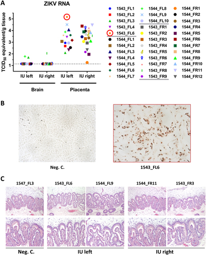
a ZIKV-specific RT-qPCR analysis of fetal brain and placenta samples. The cut-off for the PCR was 1.14 TCID50 equivalent/g tissue. Infectious virus was recovered from the placenta sample from fetus 1543_FL6 (red circle). b IPMA of Vero cells inoculated with placenta sample 1543_FL6. c Representative panel of HE-stained placenta samples derived from the IU-inoculated sows
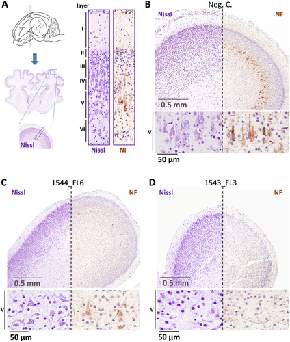
a Overview of the localization of the parietal lobe and its cytoarchitecture in the normal fetal pig brain. The different cortical layers (I–VI) are indicated. b Images of Nissl and neurofilament (NF)-stained parietal lobe cross-sections of a negative control fetus and a higher magnification of the pyramidal neurons of cortical layer V. c A fetus presenting moderate neurodepletion in cortical layer V (1544_FL6) and d a fetus presenting severe neurodepletion in cortical layer V with prominent lobe hypoplasia (1543_FL3)
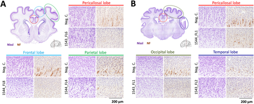
Nissl and neurofilament (NF)-stained cross-sections of the cerebral cortical lobes at the striatum a and thalamus b of other inoculated fetuses presenting neurodepletion. The various cerebral lobes are indicated. For reference, images are included which are derived from similar cross-sections and from the same cerebral lobes of negative control fetuses. NF neurofilament