Figures & data
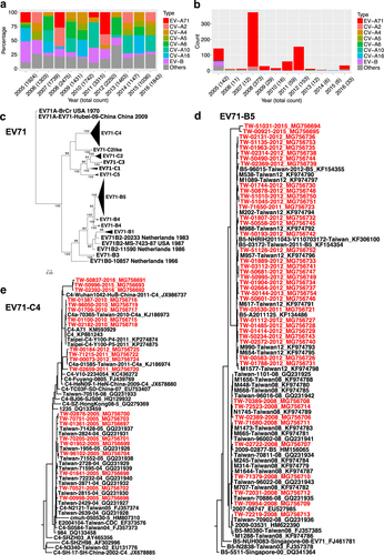
a Percentages of enterovirus infections since 2005, including EV-A71 (marked in red), six CV types (A2, A4, A5, A6 A10, and A16 in different colors), EV-B (purple), and other species (gray) in Taiwan. EV-B included CV types (B1–6 and A9) and echovirus types 3, 6, 11, 18, and 30. Counts of total cases reported in each year are also shown in parentheses. b Counts of severe complications are further summarized. c Compressed ML tree of Taiwanese strains in various subgenotypes (A, B0–5, and C1–5). Significant bootstrap support values greater than 70% are indicated at major nodes. d B5 and e C4 subtrees are shown. The tip labels of the 63 strains in B5 and C4 sequenced in this study are colored in red
Taiwanese genomes of EV-A71 analyzed in this study
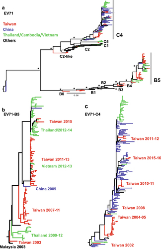
a Compressed tree, including Taiwanese and worldwide strains. Significant bootstrap support values greater than 70% are indicated by asterisks at the major nodes. Strains isolated from Taiwan, China, Cambodia/Thailand/Vietnam, and other countries are marked in different colors. b B5 and c C4 subtrees are shown
Cross-genotypic patterns are found in the compressed trees of the (a P1, b P2, and c P3) regions, rooted by the oldest prototype strain (CVA2-Fleetwood), which was isolated in 1947. Comparing to the P1 region, two reclusterings containing EV-A71 C2-like/C4 with B3 and CV-A8 with EV-A71 C in the P2 and P3 region are highlighted in blue. Through intratypic recombination, the B3 subgenotype is a potential origin of recombinants. d EV-A71 C4 and e C2-like strains were detected by SimPlot (upper panel) and Bootscan (lower panel) analyses. Reference strains, as noted, are marked in different colors. Numbers indicate nucleotide positions in the CDS corresponding to the viral genome
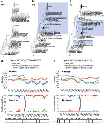
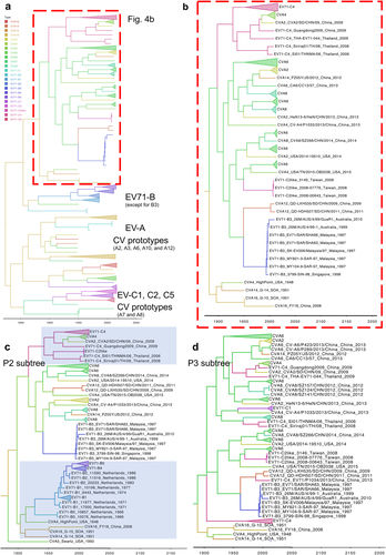
Bayesian phylogenetic trees based on (a) and b. P2/P3, c P2 and d P3 regions. Genotypes/subgenotypes of EV-A71 and CVs are marked in different colors. The subtrees in (b–d) contain mixed clusters, showing intra- and intertypic recombination events among EV-A71 B3/C4 and CVs. Highlights in c represent the reclustering of the prototype CVs with EV-A71 B and of EV-A71 C4 with the currently circulating CV-A4
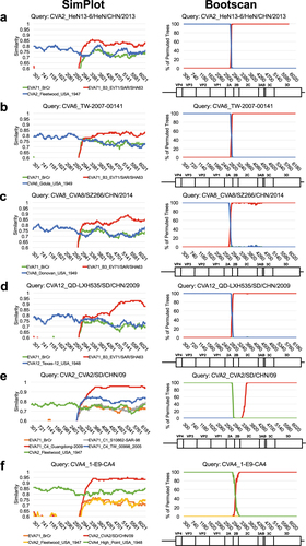
Recombination among the currently circulating CVs was examined. Using EV-A71 B3 as the reference strain, a single crossover at the P1/P2 boundary was detected by SimPlot (left panel) and Bootscan (right panel) analyses by querying (a) CV-A2, (b) CV-A6, (c) CV-A8, and d CV-A12. The roles of (e) EV-A71 C4 recombined with CV-A2 and f intertypic recombination of CV-A2 and CV-A4 are further examined. The references used in each analysis are marked in different colors, and coding nucleotide positions corresponding to the viral genome are indicated
Amino acid positions of 11 signatures carried by circulating strains of recombinant EV-A viruses
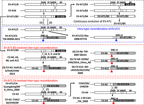
A simplified flowchart to illustrate several recombination events identified in this study. The currently circulating EV-A71 B5 strain was derived from the B4 strain, which was an intratypic recombinant virus of EV-A71 genotype B (black rectangle). The previously active EV-A71 B3 strain that carried the CV-A16-like 3D genome also exhibited intratypic recombination (blue rectangle), resulting in the emergence of C4 (#1) and C2-like strains (#2). In addition, intertypic recombination (upper red rectangle) between EV-A71 B3 and several CVs is also shown (#3–6). EV-A71 C4-related intertypic recombination occurred in ~2009 to generate a particular CV-A2 strain (CVA2/SD/CHN/09), which further recombined with CV-A4 to generate the currently circulating CV-A4 (#7 and #8, lower red rectangle). Breakpoints corresponding to the coding nucleotide positions of each recombination event are indicated