Figures & data
Eight-week-old female BALB/c mice (n = 6) were anesthetized with isoflurane and infected intranasally with 1, 10, 100, or 1000 pfu of H5N6/GZ14. The mice were monitored for 14 days. a The body weights were measured every other day. The results from each group and each time point are expressed as the mean ± standard deviations (SD). b Survival of infected mice. Mice were euthanized when body weight loss exceeded 25% of the original weight
Replication of influenza H5N6/GZ in mice
a, b Eight-week-old female BALB/c mice without infection. c–h Eight-week-old female BALB/c mice infected with 10 MLD50 (50 pfu) of H5N6/GZ14. Lung tissue sections were stained with hematoxylin-eosin and analyzed under a light microscope. At 1 dpi, inflammatory cells (arrows) could be observed around the bronchi (c, d). The airways showed small volumes of exudates with edema fluid mixed with erythrocytes and inflammatory cells (asterisks). e, f At 3f dpi, the lungs had high numbers of inflammatory cells (arrows), bronchial epithelial intracellular edema, and necrosis with necrotic epithelium sloughing into the airway spaces (asterisks). g, h At 5 dpi, severe pulmonary parenchyma consolidation was observed. Increased accumulation of inflammatory cells and necrotic tissue debris (arrows) was observed in the lung parenchyma. The alveoli were completely filled with edema and hemorrhages (asterisks). Scale bars = 100 μm (a, c, e, g) and 25 μm (b, d, f, h)
The lung tissues were collected and homogenized from H5N6/GZ14-infected mice at 1, 3, and 5 dpi (n = 3). The levels of cytokines and chemokines in the lung homogenates (pg/g of lung tissue) were determined by ELISA. The results from each time point are expressed as the mean ± SD. *A significant difference (*P < 0.05)
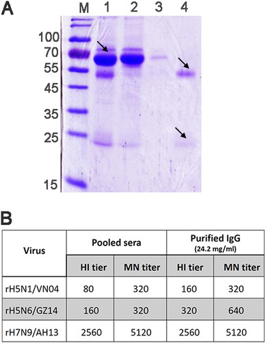
a Coomassie Blue staining after SDS-PAGE of macaque serum and purified IgG. M molecular weight markers (kDa). Lane 1: pooled hyperimmune rhesus macaque serum (diluted 500-fold). The filtered start serum mainly contains albumin (arrow). Lane 2: the flow through pool and unbound material (diluted 50-fold). Albumin and other proteins were removed and could be observed in the flow through pool. Lane 3: the effluent and column wash. Lane 4: the eluted and purified IgG (diluted 1000-fold). The IgG heavy chain (~50 kDa) and light chain (~25 kDa) (arrows) could be observed in the pooled elution buffer without albumin and other proteins. b HI and the MN neutralizing antibody titer of the pooled hyperimmune sera and the purified polyclonal IgG antibody. Three recombinant viruses, rH5N1/VN04, rH5N6/GZ14, and rH7N9/AH13, were used to immunize the rhesus macaques. HI titers are presented as the reciprocal value of the highest serum dilution that inhibited hemagglutination. MN titers are presented as the reciprocal value of the highest serum dilution that conferred 50% neutralization of 100 TCID50 of the virus
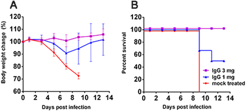
Female 8-week-old BALB/c mice (n = 6) were intranasally infected with 2 MLD50 (10 pfu) of H5N6/GZ14. One day later, the mice were treated with an intraperitoneal injection of either 1 or 3 mg of anti-influenza IgG/mouse. The mock-treated mice received an intraperitoneal injection of 3 mg of purified unrelated IgG. a The body weights were measured every other day. The results from each group and each time point are expressed as the mean ± SD. b Survival of infected mice. Mice were euthanized when body weight loss exceeded 25% of the original weight