
Figures & data
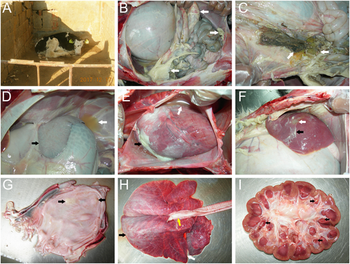
a The newborn calves showed depression, poor appetite, and paralysis. b A large number of light yellow fibrinous suppurative clots in the abdominal cavity (white arrows). c Gastric contents outflowing from an ulcer in the gastric fundus (white arrows). d Yellow effusion in the abdominal cavity (white arrows) and white fibrous protein on the surface of the spleen (black arrows). e A large amount of yellow pericardial effusion (white arrows) and white septic exudate in the epicardium (black arrows). f Liver abscessation (white arrows) with white fibrinous purulent exudate (black arrows) on the surface of the liver. g White purulent material in the bladder (black arrows). h The connective tissue hyperplasia in the anterior lung lobe (white arrows), edema in the posterior lung lobe (black arrows), and a large amount of white foamy fluid in the trachea (yellow arrows). i Yellow suppurative nodules in the renal papillae (black arrows)