Figures & data
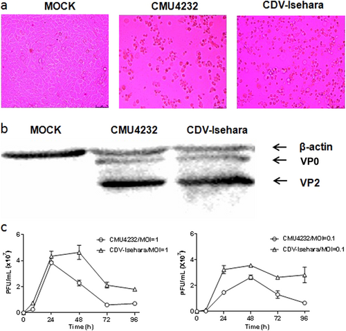
a Cytopathic effects at 1dpi displayed on RD cells infected with CMU4232 or CDV-Isehara. b Detection of viral structural proteins VP0 and VP2 of CMU4232 and CDV-Isehara by western blotting with antibody specific for EV-A71. β-actin was used as a internal control. c Growth curves of CMU4232 and CDV-Isehara in RD cells. RD cells were inoculated with CMU4232 or CDV-Isehara as indicated at either MOI of 1 or 0.1. Samples were collected at the times indicated and titrated by PFU assay. All assays were performed in triplicate. At each time point, titer values are means of three samples; error bars represent SEM. dpi days post infection, PFU plaque forming unit, SEM standard error of mean
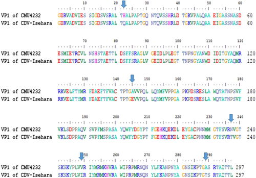
In total, there were 297 amino acids in the VP1 protein of EV-A71, and five residue variations (22 H/Q, 145 A/E, 237 N/T, 249 V/I, and 289 A/T) were found in the VP1 amino acid sequence between CMU4232 and CDV-Isehara strains
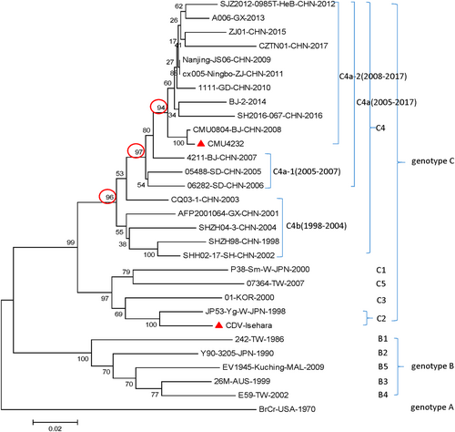
CMU4232, CDV-Isehara, 18 representative CHN strains, and 10 international representative strains of different subgenotypes are included in this dendrogram. The 18 representative CHN strains were selected from different isolation places in mainland China from 1998 to 2017 according to available data on internet. Details of all the EV-A71 strains included in the dendrogram are provided in Table S1. CHN Chinese
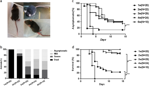
The 1, 2, 3, 4, and 6-week-old hSCARB2 Tg mice were infected intracerebral (I.C.) with CDV-Isehara strain at a dose of 4.8 × 10⁶ pfu and monitored daily after infection. a The CNS-like hindlimb paralysis, ruffled fur, and shrinking. b The clinical symptoms (asymptomatic, mild, severe, and dead), followed by the criteria described in the materials and methods section (c) asymptomatic (%) and (d) survival (%). Significant difference of asymptomatic (%) and survival (%) with different weeks old Tg mice infected with CDV-Isehara was shown as ****p,0.0001. pfu plaque forming unit, CNS central nervous system, I.C. intracerebral
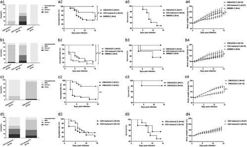
Three-week-old hSCARB2 KI mice were inoculated I.C. with DMEM/ EV-A71 (a) or inoculated intravenous (I.V.) with DMEM/EV-A71 (b) respectively. Three-week-old hSCARB2 KI mice were inoculated with CMU4232 (c) /CDV-Isehara (d) strain via I.C. or I.V. route respectively. (1) The clinical symptoms, (2) asymptomatic (%), (3) survival (%), and (4) body weight change (%) of the Tg mice were observed and recorded daily for 2 weeks after viral infection. ***p,0.000. I.C. intracerebral, I.V. intravenous
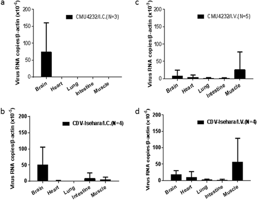
Three-week-old hSCARB2 KI mice were inoculated with CMU4232/ CDV-Isehara via I.C. or I.V. route. EV-A71 genome copy numbers in the brain, heart, lung, intestine, and muscle of EV-A71-infected Tg mice were determined at several time points by quantitative RT-PCR. Mouse β-actin gene expression in each tissue was used as the internal control. Results represent the mean of three to five samples; error bars represent STDEV. a CMU4232/I.C. (N = 3); b CDV-Isehara/I.C. (N = 4); c CMU4232 /I.V. (N = 5); d CDV-Isehara/I.V. (N = 4)
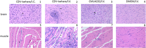
Sporadic neuronal necrosis and neuronophagia (arrowhead) in the brain (1) and Inflammatory cell infiltration (arrowhead) in the skeletal muscle (5) of mouse model with CDV-Isehara/I.C.; glial nodule (arrow) and neuronophagia (arrowhead) in the brain (2) and severe necrotizing myositis with a mass of inflammatory cell infiltration (arrow) in the skeletal muscle (6) of mouse model with CDV-Isehara/I.V.; perivascular cuffing (arrowhead), partial neuronal necrosis and neuronophagia in the brain (3) and degeneration and necrosis in muscle bundles with inflammatory cell infiltration (arrowhead) (7) in mouse model with CMU4232/I.V.; the brain (4) and the skeletal muscle (8) of MOCK mouse with DMEM/I.V. (magnification: 200 × ). Representative samples are shown for each group
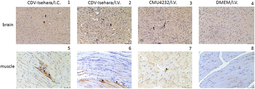
The brain (1) and the skeletal muscle (5) of mouse model with Isehara/I.C.; the brain (2) and the skeletal muscle (6) of mouse model with CDV-Isehara/I.V.; the brain (3) and the skeletal muscle (7) of mouse model with CMU0804/I.V.; the brain (4) and the skeletal muscle (8) of MOCK mouse with DMEM/I.V. Arrowheads indicate EV-A71-positive cells (IHC staining against EV-A71 VP2, magnification: 200 × ). Representative samples are shown for each group