"Single coronary artery with retrosternal course." Acta Cardiologica, 78(8), pp. 964–965
Disclosure statement
The authors declare that they have no known competing financial interests or personal relationships that could have appeared to influence the work reported in this paper.
Figure 1. Cardiac computed tomography angiography image showing dominant right coronary artery (a) and its two branches: the first vessel (b), the second one (c).
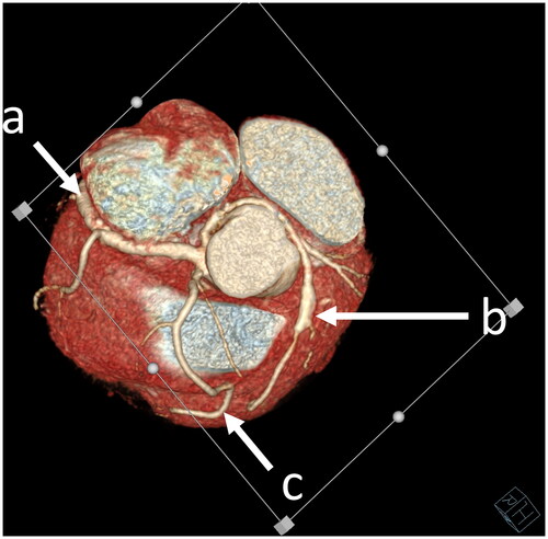
Figure 2. Another CCTA scan revealing single coronary artery(a) and two vessels (b,c) arising from the dominant one.
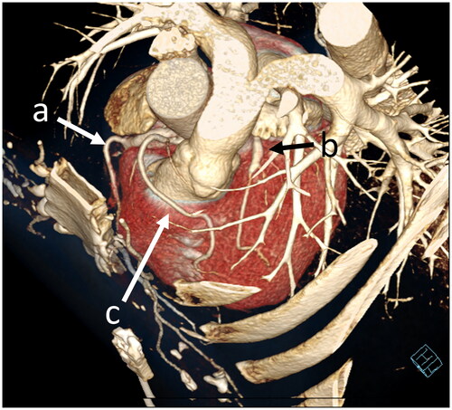
Figure 3. The last CCTA image, which shows right coronary artery(a) and two vessels (b,c) coming from the dominant one.
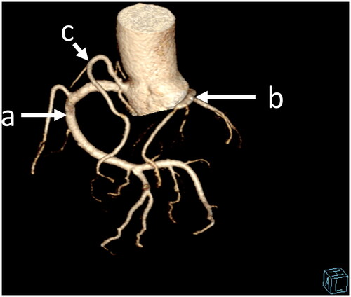
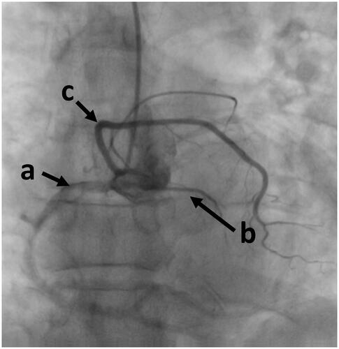
Figure 4. Coronary angiography showing dominant right coronary artery (a) and its two branches: the first vessel (b), the second one (c).