
Figures & data
Figure 1. (a–f) Sequential steps of macroscopic examination of the gallbladder by the surgeon. Full video is available in the Supplementary appendix.
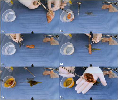
Table 1. Reasons for additional histopathologic examination.
Figure 2. (a, b) Macroscopic images of a normal appearance of the gallbladder mucosa. Please notice the smooth and glistening serosa and folded mucosa. The numerous spots are cholesterolosis, which is accumulation of lipoma filled macrophages in the mucosa. (c, d) Macroscopic image of acute cholecystitis gallbladder.
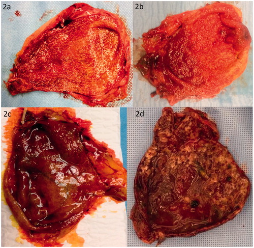
Figure 3. (a) Macroscopic image of a 1.8 cm large adenomyoma of the gallbladder at the hepatic side after 24 h of formaldehyde fixation. (b, c) Macroscopic image of a hyperplastic polyp is indicated with arrow (◄). (d, e) Macroscopic image of a multiple cholesterol gallbladder polyps indicated with arrows (◄).
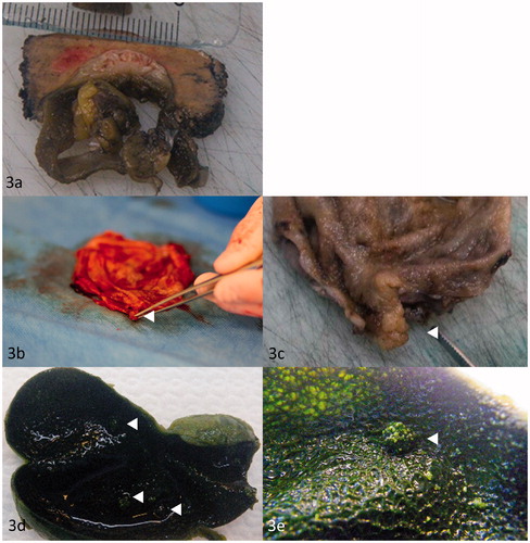
Figure 4. (a, b) Macroscopic image of a porcelain gallbladder. (c) Macroscopic image of gallbladder containing T2 GBC in the fundus of the gallbladder. Mucosal irregularity containing GBC is indicated with arrow (◄). (d, e) Macroscopic image of gallbladder containing T3 GBC in the fundus of the gallbladder (◄) and liver metastasis (◄◄). (f) Macroscopic image of gallbladder with liver resection (◄). (g) Macroscopic image of intracholecystic papillary neoplasm with highgrade dysplasia (◄◄).
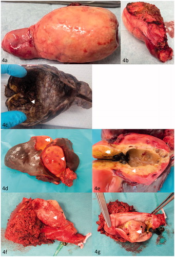
Table 2. Number of cholecystectomies per year and the number of histopathologic gallbladder specimens sent to department of pathology.