
Figures & data
Figure 1. (A) CT-scan showing the nephrolithiasis in the renal allograft. (B) Endoscopic image of the stone before and after opening the mucosa. (C) Set-up of the renal allograft on ice.
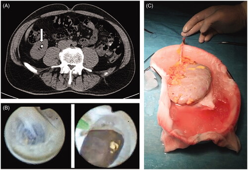
Table 1. Literature overview for back-table endoscopic treatment of nephrolithiasis in renal allografts.