
Figures & data
Table 1. Basic data and occlusal characteristics of Osteogenesis imperfecta (OI) patient sample and healthy age- and gender-matched controls.
Figure 1. Dental arch measurements. 1. Upper arch length from the extreme labial surfaces of the incisors perpendicular to a line connecting the extreme mesial points of the first permanent molars. 2. Upper inter-canine width, between the crown tips. 3. Upper inter-premolar widths, between the palatal cusp tips of both first and second premolars. 4. Upper inter-molar width, between the mesiopalatal cusp tips of first permanent molars. Similarly: 5. Lower arch length, 6. Lower inter-canine width, 7. Lower inter-premolar widths, 8. Lower inter-molar width.
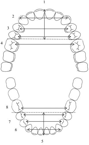
Figure 2. Palatal height measurements. (A) The black line marked B indicates the cross-section level in (B), and the white line marked C indicates the cross-section level in (C). B) Anterior palatal height measured from the mid-palate (1) to two reference lines: a line (2) drawn at the gingival margin level of the first premolars, and a line (3) on their palatal cusp tips. C) Posterior palatal height measured from the mid-palate (4) to two reference lines: a line (5) drawn at the gingival margin level of the first permanent molars, and a line (6) on their mesiopalatal cusp tips.
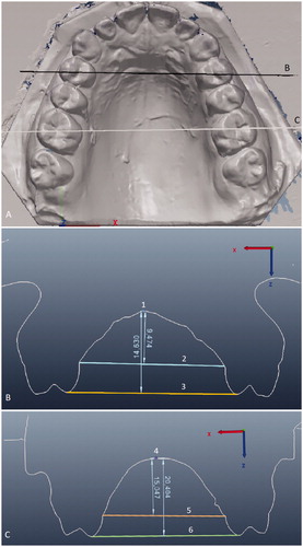
Table 2. Mesio-distal widths of permanent teeth, third molars excluded, in patients with Osteogenesis imperfecta (OI) and in controls.
Table 3. Dental arch dimensions of patients with Osteogenesis imperfecta (OI) and healthy controls; means (and standard deviation) in millimetres.
Table 4. Palatal height of patients with Osteogenesis imperfecta (OI) and healthy controls; means (and standard deviation) in millimetres.
Data availability statement
The authors confirm that the data supporting the findings of this study are available within the article. Raw data were generated at University of Helsinki and is available from the corresponding author HA on request.