Figures & data
Figure 1. Desquamated epithelial cells of human whole saliva contain hCAP18/LL-37 protein. Desquamated oral epithelial cells treated with exogenous LL-37 (1 µM) for 4 h (+) contain about 30 times more hCAP18/LL-37 than non-treated control cells (−). Desquamated oral epithelial cells were isolated from all four volunteers enrolled in the study, and hCAP18/LL-37 protein levels were analyzed in samples from each of the four individuals. Values are presented as mean ± standard error of the mean of four individuals in each group. ***represents p < .001.
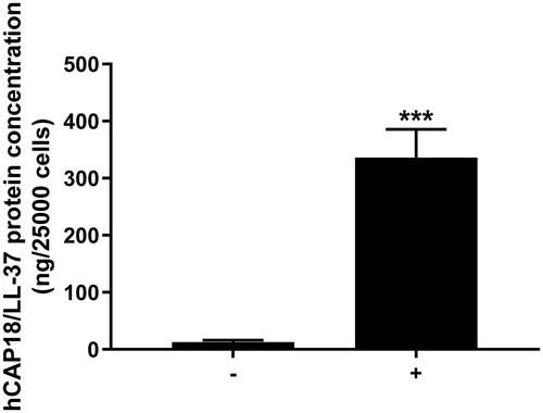
Figure 2. Desquamated oral epithelial cells spontaneously release/leak hCAP18/LL-37. Desquamated oral epithelial cells were isolated from three of the four volunteers included in the study and incubated in PBS for 20, 70 and 130 min, and at the end of incubation cells were removed and the supernatant collected for analysis. The levels of hCAP18/LL-37 in the medium were determined for each subject and time point. No hCAP18/LL-37 was observed in non-conditioned PBS, whereas PBS which had been in contact with cells for 20, 70 and 130 min contained the peptide. Data are presented as mean ± standard error of the mean of three individuals. * and ** represent p < .05 and p < .01, respectively, vs. data for the 20 min time point.
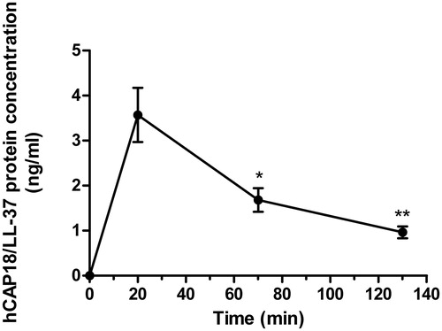
Figure 3. Incubation with synthetic LL-37 for 24 h reduces S. mutans viability. S. mutans (A-C) or S. gordonii (D-F) were treated with 1 (B, E) or 8 (C, F) µM LL-37 and the proportion of live and dead bacteria determined. Controls (A, D) received vehicle as appropriate. Experiments were performed in duplicate, and images are representative for each group. Scale bar in panel F represents 25 µm (for all images).
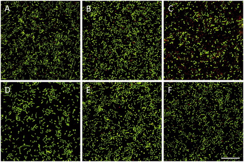
Figure 4. Incubation with synthetic LL-37 but not homogenates of desquamated oral epithelial cells for 24 h attenuates S. mutans viability. Neither cellular homogenates prepared from 100,000 cells per ml PBS (B) nor homogenates from 500,000 cells per ml PBS (C) had any impact on number of live and dead bacteria compared to control (A). Summarized data are shown in panel F. Also, incubation with homogenate of cells (500,000 cells per ml PBS) pre-treated with 1 µM LL-37 for 4 h to spike them with LL-37 had no effect on S. mutans viability (D, F), whereas synthetic LL-37 (8 µM), included as positive control, reduced the number of live bacteria by about 40% (E, F). Cells were isolated from one of the volunteers included in the study, and experiments were performed as independent experiments in duplicate. All experiments were performed with cells isolated from the same individual. For each sample, 10 images were analyzed, and the number of live bacteria presented as percent of total number. Values are presented as mean ± standard error of the mean of 9–10 observations in each group. *** represents p < .001 vs. control. Scale bar in panel E represents 25 µm (for all images).
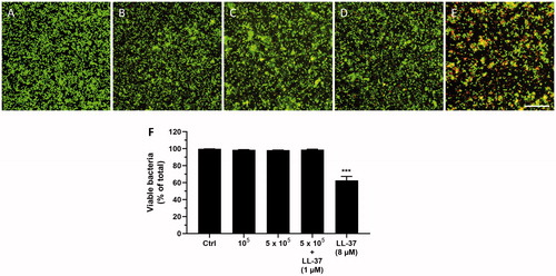
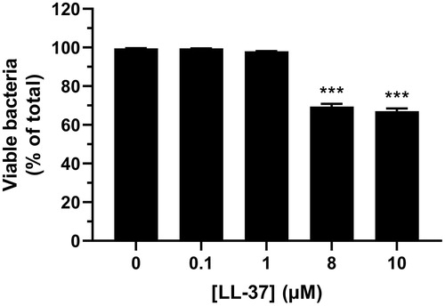
Figure 5. Treatment with 8 and 10 µM of synthetic LL-37 reduces S. mutans viability. Bacteria were treated with or without LL-37 (0.1, 1, 8 and 10 µM) for 24 h and the proportion of live and dead bacteria assessed. Experiments were performed in triplicate. Values are presented as mean ± standard error of the mean of 29–30 observations in each group. *** represents p < .001 vs. control.