Figures & data
Table 1. Summary of the six patient cases included in the survey.
Figure 1. Example of a patient case included in the survey.
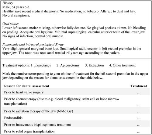
Figure 2. Model for grouping of consensus on the choice of dental treatment. High consensus: ≥75% of participants agreed on choice of dental treatment. Moderate consensus: 50–74% of participants agreed on choice of dental treatment. Low consensus: <50% of participants agreed on choice of dental treatment. Treatments A–D different for each patient case.
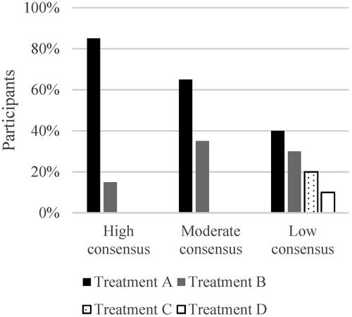
Figure 3. Model for grouping of variability of the choice of dental treatment. High variability: ≥50% of participants altered their treatment selection in relation to underlying medical issue. Moderate variability: 25–49% of participants altered their treatment selection in relation to underlying medical issue. Low variability: <25% of participants altered their treatment selection in relation to underlying medical issue.
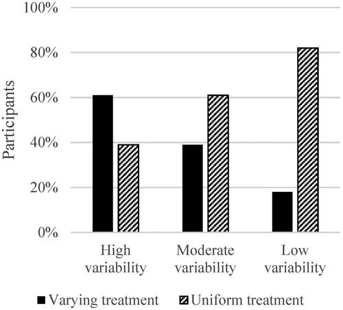
Figure 4. Flow-chart of study participation. w: Women; m: men.
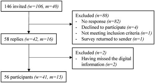
Figure 5. Estimation of the importance of dental evaluations in relation to the various medical situations. HVS: Heart valve surgery; CT: chemotherapy; RT: radiation therapy; EC: endocarditis; IVBP: intravenous bisphosphonate treatment; SOT: solid organ transplantation.
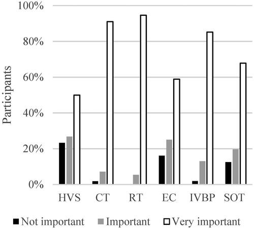
Figure 6. Views on the level of scientific evidence for performing dental evaluations in relation to the various medical situations. HVS: Heart valve surgery; CT: chemotherapy; RT: radiation therapy; EC: endocarditis; IVBP: intravenous bisphosphonate treatment; SOT: solid organ transplantation.
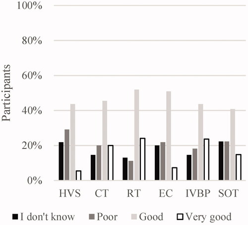
Figure 7. Estimation of risk for complications if patients in cases 1–6 were to be left without dental treatment in relation to the various medical situations. HVS: Heart valve surgery; CT: chemotherapy; RT: radiation therapy; EC: endocarditis; IVBP: intravenous bisphosphonate treatment; SOT: solid organ transplantation.
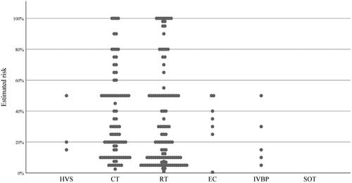
Figure 8. Correlation between estimated risk for complications and rate of extractions. Y-axis: proportion of estimations in each case where extraction was selected. X-axis: estimated risk for complications. Each dot represents a case assessed by a participant having defined the risk in percentage.
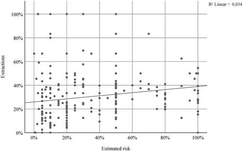
Figure 9. Relation between estimated risk for complications and participant years of experience of working with hospital-affiliated dentistry. Each dot represents a case assessed by a participant having defined the risk in percentage.
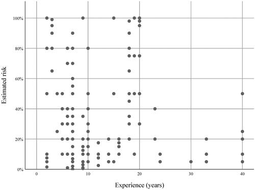
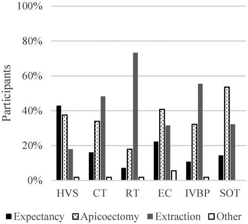
Figure 10. Case 2: Proposed treatment for an asymptomatic root canal treated upper left second premolar with apical radiolucency in various medical situations. HVS: Heart valve surgery; CT: chemotherapy; RT: radiation therapy; EC: endocarditis; IVBP: intravenous bisphosphonate treatment; SOT: solid organ transplantation. Other treatment unspecified by participants.