1,447
Views
8
CrossRef citations to date
0
Altmetric
Original Article

Airtraq laryngoscope: a solution for difficult laryngeal exposure in phonomicrosurgery

, , , , &
Pages 635-639 | Received 17 Oct 2016, Accepted 23 Nov 2016, Published online: 13 Jan 2017

Figures & data

Table 1. Clinical features of patients, possible causes of difficult laryngeal exposure (DLE), and grade of laryngeal exposure.

Figure 1. Airtraq® laryngoscope has an anatomically shaped blade and can be connected with a video camera system (A). A set of curved instruments designed by our team (B). In operation, Airtraq® was connected with an endoscopic video system and, thus, the images of the larynx were displayed on a monitor. The surgeon held Airtraq® with the left hand and the instrument with the right hand (C).

Figure 1. Airtraq® laryngoscope has an anatomically shaped blade and can be connected with a video camera system (A). A set of curved instruments designed by our team (B). In operation, Airtraq® was connected with an endoscopic video system and, thus, the images of the larynx were displayed on a monitor. The surgeon held Airtraq® with the left hand and the instrument with the right hand (C).

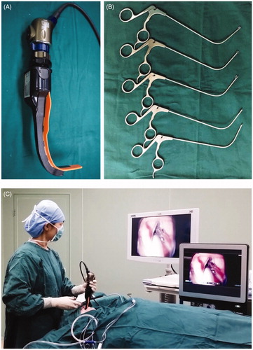
Figure 2. Laryngeal exposure with the conventional suspension laryngoscope (Case No. 5 in ). With the conventional suspension laryngoscope, only the arytenoid area could be viewed microscopically, as grade 3 laryngeal exposure.

Figure 2. Laryngeal exposure with the conventional suspension laryngoscope (Case No. 5 in Table 1). With the conventional suspension laryngoscope, only the arytenoid area could be viewed microscopically, as grade 3 laryngeal exposure.

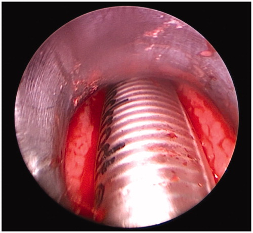
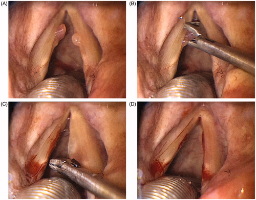
Figure 3. Laryngeal exposure with Airtraq® laryngoscope (Case No. 5 in ). When applying the Airtraq® laryngoscope, the entire glottis was exposed including the anterior commissure, as grade 1 laryngeal exposure. The bilateral polys were also visualized clearly (A). The left vocal cord polyp was grasped with the special curved cutting forceps (B). The right vocal cord polyp was removed successfully by the same method (C). The vocal cord polyps were just removed completely (D).

Figure 3. Laryngeal exposure with Airtraq® laryngoscope (Case No. 5 in Table 1). When applying the Airtraq® laryngoscope, the entire glottis was exposed including the anterior commissure, as grade 1 laryngeal exposure. The bilateral polys were also visualized clearly (A). The left vocal cord polyp was grasped with the special curved cutting forceps (B). The right vocal cord polyp was removed successfully by the same method (C). The vocal cord polyps were just removed completely (D).