Publication Cover
Caryologia
International Journal of Cytology, Cytosystematics and Cytogenetics
Volume 66, 2013 - Issue 3
1,584
Views
17
CrossRef citations to date
0
Altmetric
Articles

Cytogenetic effects of chitosan-capped silver nanoparticles in the Allium cepa test

Pages 275-281 | Published online: 08 Nov 2013

Figures & data

Figure 1. (Color online) Atomic force microscopy (AFM) image of AgNP.

Figure 1. (Color online) Atomic force microscopy (AFM) image of AgNP.

Figure 2. (Color online) Stages of mitosis in the meristematic cells of A. cepa: (a) prophase; (b) metaphase; (c) anaphase; (d) telophase.

Figure 2. (Color online) Stages of mitosis in the meristematic cells of A. cepa: (a) prophase; (b) metaphase; (c) anaphase; (d) telophase.

Table 1. Data on mitotic and phase indexes (mean ± SD) in the meristematic cells of A. cepa roots treated with solutions of silver nanoparticles.

Figure 3. (Color online) Control meristematic cells of A. cepa.

Figure 3. (Color online) Control meristematic cells of A. cepa.

Table 2. Data on the number of mitotic disturbances (mean ± SD) in the meristematic cells of A. cepa roots treated by AgNP.

Figure 5. (Color online) Giant polyploid cells (prophases, telophases and interphases) in root meristem of A. cepa after incubation in solution of 50 mg l−1 AgNP.
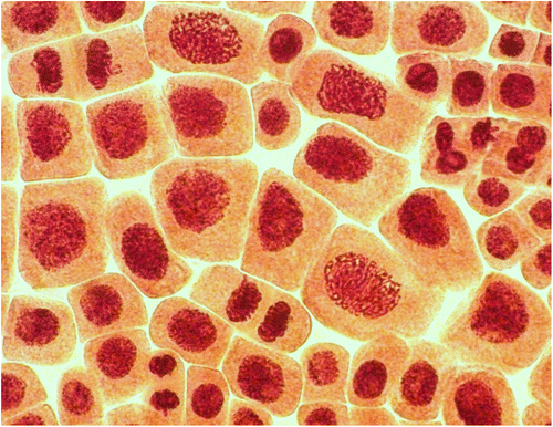
Figure 5. (Color online) Giant polyploid cells (prophases, telophases and interphases) in root meristem of A. cepa after incubation in solution of 50 mg l−1 AgNP.
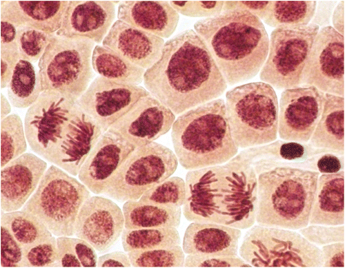
Figure 4. (Color online) Giant polyploid cells (anaphases and interphases) in root meristem of A. cepa after incubation in solution of 50 mg l−1 AgNP.

Figure 4. (Color online) Giant polyploid cells (anaphases and interphases) in root meristem of A. cepa after incubation in solution of 50 mg l−1 AgNP.

Table 3 Data on the number of chromosomal aberrations and micronuclei (mean ± SD) in the meristematic cells of A. cepa roots treated with colloidal solutions of silver nanoparticles.

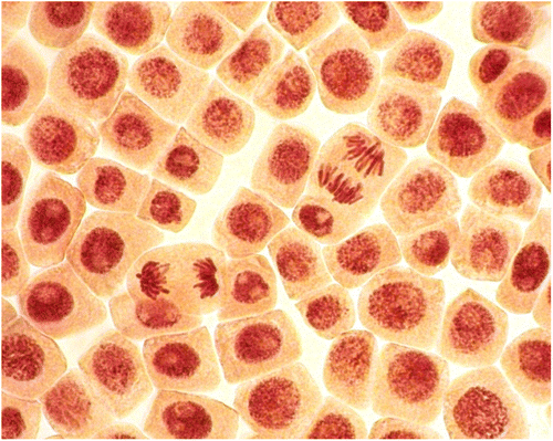
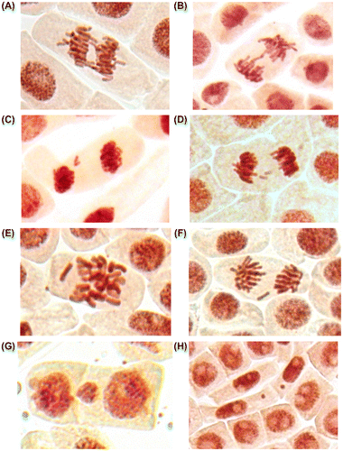
Figure 6. (Color online) Mitotic and chromosome abnormalities and micronuclei in root meristematic cells of A. cepa: bridges (A, B), acentric fragment (C), double fragments (D), lagging chromosomes (E, F) and micronuclei (G, H).

Figure 6. (Color online) Mitotic and chromosome abnormalities and micronuclei in root meristematic cells of A. cepa: bridges (A, B), acentric fragment (C), double fragments (D), lagging chromosomes (E, F) and micronuclei (G, H).

Reprints and Corporate Permissions

Please note: Selecting permissions does not provide access to the full text of the article, please see our help page How do I view content?

To request a reprint or corporate permissions for this article, please click on the relevant link below:

Academic Permissions

Please note: Selecting permissions does not provide access to the full text of the article, please see our help page How do I view content?

Obtain permissions instantly via Rightslink by clicking on the button below:

If you are unable to obtain permissions via Rightslink, please complete and submit this Permissions form. For more information, please visit our Permissions help page.