Figures & data
Figure 1. (Color online) Microsporogenesis in Fritillaria stribrnyi stained with acetic-orcein. (a) Microspore mother cells; (b) leptotene; (c) zygotene; (d) pachytene; (e) diplotene; (f) diakinesis; (g) metaphase I; (h) anaphase I; (i) telophase I; (j) dyad; (k) prophase II; (l) metaphase II; (m) anaphase II; (n) telophase II; (o) isobilateral tetrad. Scale bars = 20 μm.
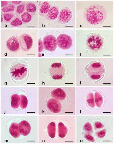
Figure 2. (Color online) Abnormalities in Fritillaria stribrnyi during microsporogenesis. (a) Lagging chromosome in metaphase I (arrow); (b) lagging chromosome in telophase I (arrow); (c, d) asynchrony in the stages of meiosis II. Scale bars = 20 μm.
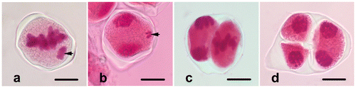
Figure 3. (Color online) Microgametogenesis in Fritillaria stribrnyi.(a) Microspore just released from the tetrad; (b) microspore with developed exine and aperture; (c) vacuolated microspore; (d) prophase; (e) metaphase; (f) anaphase; (g) telophase; (h) phragmoplast formation at the end of telophase (arrow); (i) vegetative and generative cells in pollen grain (arrow shows the wall of generative cell) (gc, generative cell; v, vacuole; vc, vegetative cell). Scale bars = 20 μm.
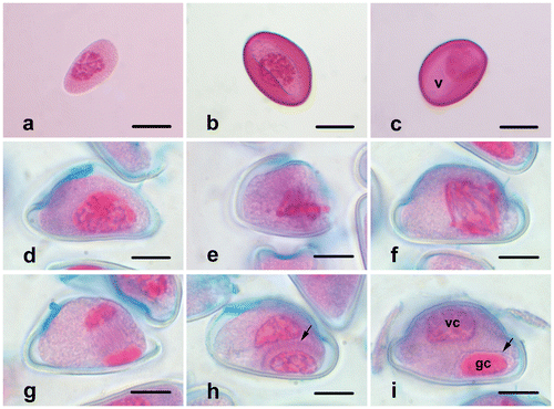
Figure 4. (Color online) Fertile and sterile pollen grains of Fritillaria stribrnyi stained with lactophenol-aniline blue solution (fp, fertile pollen; sp, sterile pollen). Scale bar = 50 μm.
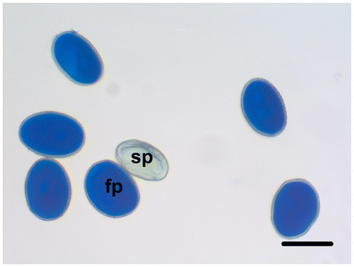
Figure 5. (Color online) Pollen tubes germinated at 25°C for 24 h and stained with lactophenol-aniline blue solution. Scale bar = 200 μm.
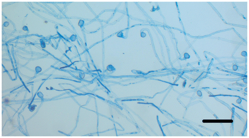
Figure 6. (Color online) Generative and vegetative nuclei stained with acetic-orcein in germinated pollen tube (gn, generative nucleus; vn, vegetative nucleus). Scale bar = 50 μm.
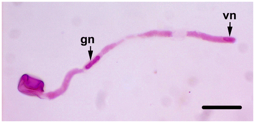
Figure 7. (Color online) Pollen tube abnormalities of Fritillaria stribrnyi. (a) Swollen pollen tube tip; (b) branched pollen tube tips. Scale bars = 20 μm.
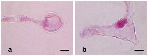
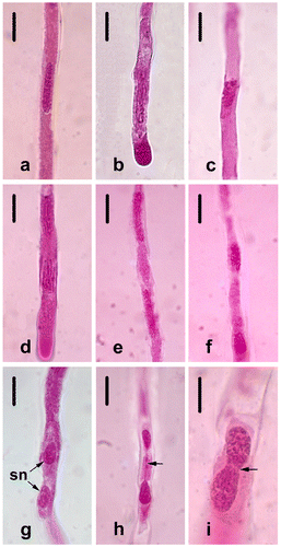
Figure 8. (Color online) Spermatogenesis of Fritillaria stribrnyi in in vitro germinated pollen tube. (a) Prophase; (b) prometaphase; (c) metaphase; (d) anaphase; (e) telophase; (f) the end of telophase; (g) two sperm nuclei; (h) telophase bridge (arrow); (i) telophase bridge (arrow) Scale bars: (a–h) 20 μm; (i) 10 μm.