Figures & data
Table 1. Restriction enzymes and chromosome digestion conditions.
Figure 1. Giemsa-stained karyotypes of the burbot, Lota lota after C-banding (a) and digestion with the restriction endonucleases DdeI (b), AluI (c) and HinfI (d); m: metacentric chromosomes, sm: submetacentric chromosomes, st-a: subtelo-acrocentric chromosomes. Scale bar = 10 μm.
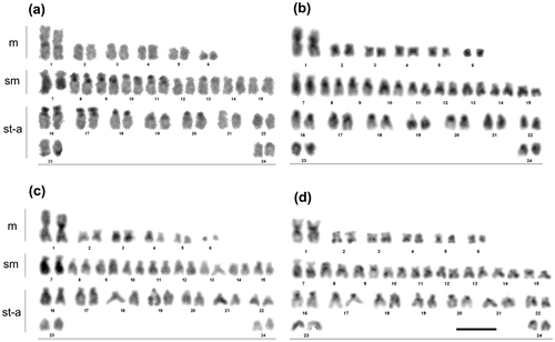
Table 2. Chromosomal distribution of C-positive heterochromatin and regions resistant to restriction endonucleases in the burbot.
Figure 2. DAPI-stained karyotype of the burbot Lota lota. The bottom row shows the NOR-bearing chromosome pair (no. 19) after sequential DAPI/chromomycin A3/silver nitrate staining. Scale bar = 10 μm.
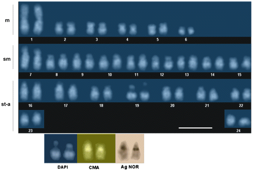
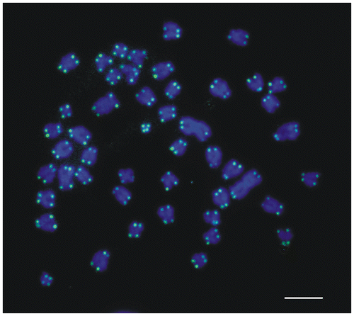
Figure 3. FISH mapping of 28S (arrows) and 5S (arrowheads) rDNA clusters to metaphase chromosomes of the burbot Lota lota stained with DAPI. Scale bar = 10 μm.
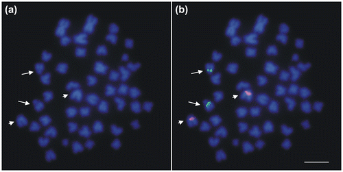
Figure 4. FISH mapping of telomeric sequences to chromosomes of the burbot Lota lota stained with DAPI. Scale bar = 10 μm.