Figures & data
Figure 1. Pollen mother cells of H. dulcis in meiotic division. (A) diakinesis with 12 bivalents; (B) diakinesis with arrow showing the chromosome associated with the nucleolus; (C) metaphase I with n = 12; (D) pollens stained with Alexander (Citation1980) staining with arrows showing unviable ones. Bar = 5 μm.
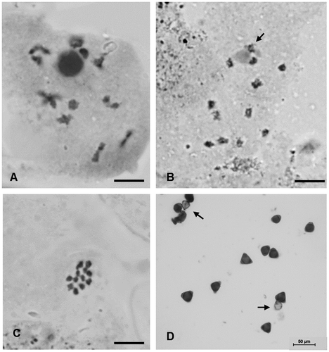
Table 1. Karyotype data of Hovenia dulcis, with average lengths of each pair of chromosomes (CL), their long arms (LA) and short arms (SA), centromeric index (CI) and chromosome classification (CC).
Figure 2. Cells of H. dulcis in metaphasic mitotic division. (A) Giemsa stained cell with 2n = 24 with arrow showing satellite; (B) DAPI stained cell with arrow showing satellite; (C) CMA stained cell with arrows showing positive bands; (D) cell counterstained with DAPI hybridized with pTa71 probe with arrows showing 45 rDNA sites; (E) cell hybridized with pScT7 probe with arrows showing 5S rDNA sites. Bar = 5 μm.
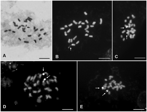
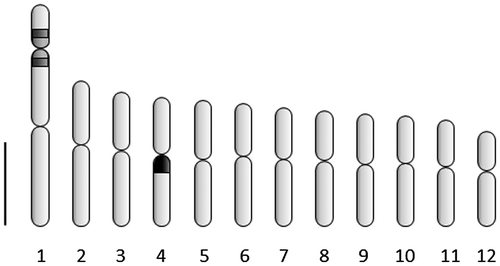
Figure 3. Idiogram of H. dulcis showing average morphological data of 12 chromosome pairs, with marks representing CMA+band (light gray), 45S rDNA site (dark gray) and 5SrDNA (black). Bar = 1 μm.