701
Views
19
CrossRef citations to date
0
Altmetric
Original Articles

New acritarch species from Holocene sediments in central West Greenland

Pages 81-88 | Received 12 May 2003, Accepted 27 Oct 2003, Published online: 06 Oct 2011

Figures & data

Location of the coring stations in Disko Bugt, central West Greenland (modified from Kuijpers et al. Citation2000).
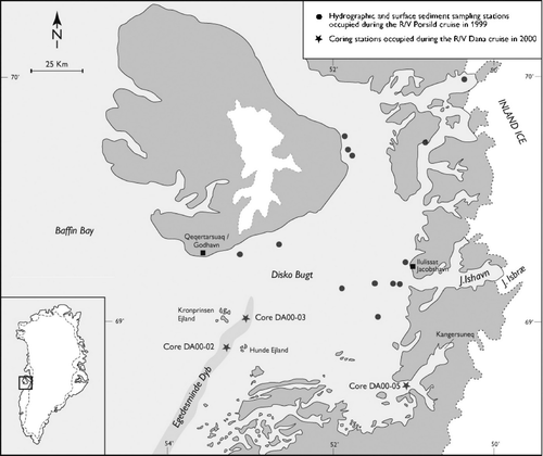
Location of the coring stations in Disko Bugt, central West Greenland (modified from Kuijpers et al. Citation2000).

A–L. Palaeostomocystis fritilla (Bujak Citation1984) comb. nov. from core DA00-03, Egedesminde Dyb, central West Greenland. The same scale is used for all specimens except (L). A. MGUH 26853. Semilateral view in high focus. Note the undulate pylome margin. B. Same specimen as (A). Semilateral view in sectional focus. C. MGUH 26855. Lateral view in sectional focus. Damaged specimen with operculum in the interior of the vesicle. D. MGUH 26856. Lateral view in high focus. E. MGUH 26858. Lateral view in sectional focus. F. MGUH 26854. Lateral view in low focus. Note the thick vesicle wall. G. Same specimen as (F). Lateral view in high focus. Note the faveolate wall structure. H. Same specimen as (D). Lateral view in low focus. I. MGUH 26857. Lateral view in low focus. J. Same specimen as (I). Lateral view in sectional focus. K. Same specimens as (I). Lateral view in high focus. L. Same specimen as (I). Faveolate wall structure in high magnification. The faveoli internal part is in focus in the centre of the picture; the reticulate muri network is shown on the periphery of the picture. Scale bars – 15 μm (A – K); 5 μm (L).
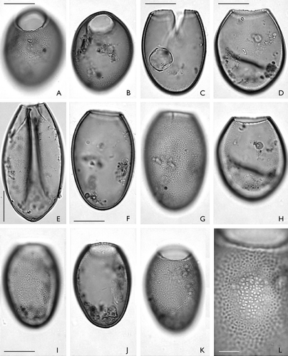
A–L. Palaeostomocystis fritilla (Bujak Citation1984) comb. nov. from core DA00-03, Egedesminde Dyb, central West Greenland. The same scale is used for all specimens except (L). A. MGUH 26853. Semilateral view in high focus. Note the undulate pylome margin. B. Same specimen as (A). Semilateral view in sectional focus. C. MGUH 26855. Lateral view in sectional focus. Damaged specimen with operculum in the interior of the vesicle. D. MGUH 26856. Lateral view in high focus. E. MGUH 26858. Lateral view in sectional focus. F. MGUH 26854. Lateral view in low focus. Note the thick vesicle wall. G. Same specimen as (F). Lateral view in high focus. Note the faveolate wall structure. H. Same specimen as (D). Lateral view in low focus. I. MGUH 26857. Lateral view in low focus. J. Same specimen as (I). Lateral view in sectional focus. K. Same specimens as (I). Lateral view in high focus. L. Same specimen as (I). Faveolate wall structure in high magnification. The faveoli internal part is in focus in the centre of the picture; the reticulate muri network is shown on the periphery of the picture. Scale bars – 15 μm (A – K); 5 μm (L).

A–L. Palaeostomocystis subtilitheca sp. nov. from core DA00-03, Egedesminde Dyb, central West Greenland. Note that the operculum occurs in the interior of each specimen except G. The same scale is used for all specimens. A. Holotype. MGUH 26841. Apical view in high focus. Note the pylome rim. B. Same specimen as (A). Apical view in sectional focus. C. Same specimen as (A). Apical view in low focus. D. MGUH 26842. Semilateral view, sectional focus. E. Same specimen as (D). Semilateral view in low focus. F. MGUH 26843. Apical view in low focus. G. MGUH 26845. Antapical view in high focus. H. MGUH 26846. Antapical view in high focus. Note small pseudopylome at 2.00 o'clock. I. Same specimens as (H). Antapical view in low focus. J. MGUH 26847. Apical view in high focus. Specimen partly damaged by lateral compression. K. Same specimen as (J). Apical view in low focus. L. MGUH 26851. Apical view in low focus. Note dome-shaped operculum in the bottom of the vesicle. Scale bar – 15 μm.
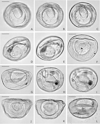
A–L. Palaeostomocystis subtilitheca sp. nov. from core DA00-03, Egedesminde Dyb, central West Greenland. Note that the operculum occurs in the interior of each specimen except G. The same scale is used for all specimens. A. Holotype. MGUH 26841. Apical view in high focus. Note the pylome rim. B. Same specimen as (A). Apical view in sectional focus. C. Same specimen as (A). Apical view in low focus. D. MGUH 26842. Semilateral view, sectional focus. E. Same specimen as (D). Semilateral view in low focus. F. MGUH 26843. Apical view in low focus. G. MGUH 26845. Antapical view in high focus. H. MGUH 26846. Antapical view in high focus. Note small pseudopylome at 2.00 o'clock. I. Same specimens as (H). Antapical view in low focus. J. MGUH 26847. Apical view in high focus. Specimen partly damaged by lateral compression. K. Same specimen as (J). Apical view in low focus. L. MGUH 26851. Apical view in low focus. Note dome-shaped operculum in the bottom of the vesicle. Scale bar – 15 μm.

A–I. Palaeostomocystis subtilitheca sp. nov. from core DA00-03, Egedesminde Dyb, central West Greenland. The same scale is used for all specimens. A. MGUH 26844. Lateral view in high focus. Specimen partly damaged by vertical compression. B. Same specimen as (A). Lateral view in low focus. Note flat operculum in the interior of the vesicle. C. MGUH 26849. Lateral view in sectional focus. Note single, solid and blunt horn at the pole diametrically opposite the pylome. D. Paratype 1. MGUH 26848. Lateral view in high focus. Note the well-developed pylome rim and the dome-shaped operculum within the vesicle. E. Same specimen as (D). Lateral view in sectional focus. F. Same specimen as (D). Lateral view in low focus. G. MGUH 26852. Antapical view in sectional focus. H. Same specimen as (G). Antapical view in low focus. I. Paratype 2. MGUH 26850. Apical view in sectional focus. Scale bar – 15 μm.
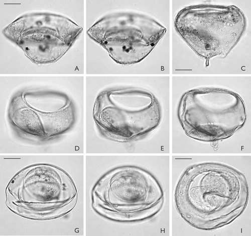
A–I. Palaeostomocystis subtilitheca sp. nov. from core DA00-03, Egedesminde Dyb, central West Greenland. The same scale is used for all specimens. A. MGUH 26844. Lateral view in high focus. Specimen partly damaged by vertical compression. B. Same specimen as (A). Lateral view in low focus. Note flat operculum in the interior of the vesicle. C. MGUH 26849. Lateral view in sectional focus. Note single, solid and blunt horn at the pole diametrically opposite the pylome. D. Paratype 1. MGUH 26848. Lateral view in high focus. Note the well-developed pylome rim and the dome-shaped operculum within the vesicle. E. Same specimen as (D). Lateral view in sectional focus. F. Same specimen as (D). Lateral view in low focus. G. MGUH 26852. Antapical view in sectional focus. H. Same specimen as (G). Antapical view in low focus. I. Paratype 2. MGUH 26850. Apical view in sectional focus. Scale bar – 15 μm.

A–E. Variation in the overall shape of Palaeostomocystis subtilitheca sp. nov. seen in lateral view. The operculum is indicated by a dashed line. The specimens are in the same scale. A. Paratype 1, illustrated on D–F. D. Illustrated on C. E. Illustrated on A−B. Scale bar – 15 μm.
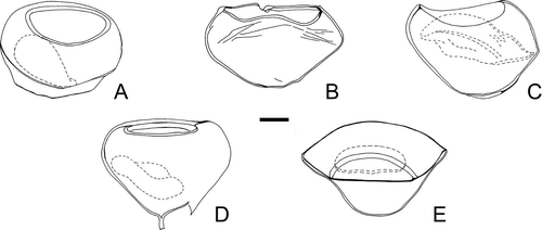
A–E. Variation in the overall shape of Palaeostomocystis subtilitheca sp. nov. seen in lateral view. The operculum is indicated by a dashed line. The specimens are in the same scale. A. Paratype 1, illustrated on Fig. 4 D–F. D. Illustrated on Fig. 4 C. E. Illustrated on Fig. 4 A−B. Scale bar – 15 μm.

Reprints and Corporate Permissions

Please note: Selecting permissions does not provide access to the full text of the article, please see our help page How do I view content?

To request a reprint or corporate permissions for this article, please click on the relevant link below:

Academic Permissions

Please note: Selecting permissions does not provide access to the full text of the article, please see our help page How do I view content?

Obtain permissions instantly via Rightslink by clicking on the button below:

If you are unable to obtain permissions via Rightslink, please complete and submit this Permissions form. For more information, please visit our Permissions help page.