Figures & data
Figure 1 A–H. Bright field micrographs of Consolea picardae anther development. A–E. Male‐fertile anther (staminate flower) cross sections. F–H. Male‐sterile anther (pistillate flower) cross sections. A. Stage 1, anther primordium with four anther wall layers: epidermis (e), endothecium (en), middle layer (ml) and tapetum (tp) surrounding the sporogenous tissue (sp). B. Early stage 2, microspore mother cell (mmc) differentiated and surrounded by four anther wall layers, e, en, ml, and binucleate tp. C. Late stage 2, meiosis is complete and a tetrahedral microspore tetrad is surrounded by callose (c); the anther wall is composed of three layers, e, en, and very active tp; ml is flattened and crushed. D. Stage 3, released and enlarged microspores (m), with large central vacuole and peripheral nucleus against the cell wall; the anther wall is still composed by three layers, e, en and tp. E. Stage 4, mature pollen filled with starch grains, the anther wall has only two remaining layers, e and en with fibrous thickenings. F. Early stage 2, mmc differentiated and anther wall with four layers, e, en, ml and radially enlarged tp. G. Stage 2, mmc degenerating and oddly shaped due to tp invasion of the anther locule; the tp layer is greatly enlarged and its cells have a single large vacuole; e, en and ml are not enlarged. H. Degenerate anther, mmc's no longer visible; the anther wall is composed of e, somewhat enlarged en, and greatly radially enlarged ml; the tp has already been crushed. Scale bars – 50 µm.
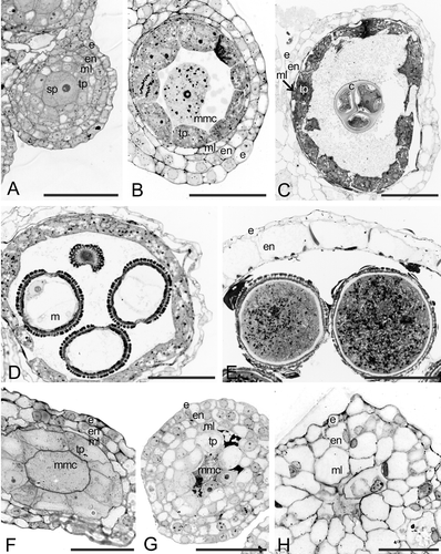
Figure 2 A–G. Transmission electron micrographs (TEM) of Consolea picardae male‐fertile anther development. A–B. Stage 1, anther primordium. A. Four young anther wall layers: epidermis (e), endothecium (en), middle layer (ml) and tapetum (tp) surrounding the sporogenous (sp) cell. B. Detail of sp cell: note very dense cytoplasm, small vacuoles (v), and abundant mitochondria and rER. C–E. Stage 2. C. Anther wall composed of three layers, e, en, flattened, crushed ml, and metabolically active tp. D. Detail of tp cell fused nuclei, showing the characteristic lobed nucleus (ln). E. Detail of tp cell cytoplasm with rER (solid arrow) and pro Ubisch bodies (hollow arrow). F–G. Stage 3: microspore. F. Detail of tp cell with dense cytoplasm and lipid bodies (lp), in which only the rER cisternae matrix is discernible (solid arrow); young Ubisch bodies are attached to the plasma membrane (hollow arrow). G. Microspore with a well‐developed exine. Scale bar – 2 µm (A, C, D, G); 1 µm (B); 0.4 µm (E, F).
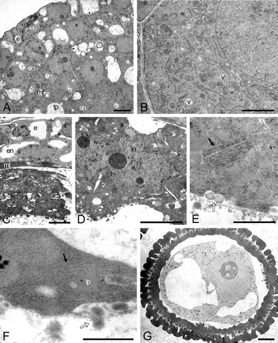
Figure 3 A–C. TEM micrographs of Consolea picardae male‐fertile mature anther and pollen grain. A. Anther wall composed of epidermis (e) and endothecium (en) with fibrous thickenings (arrows), note Ubisch bodies along lower right. B. Detail of mature pollen grain, the cytoplasm is filled with starch grains (sg) and the pollen grain wall is formed by intine (in) and exine (ex); the ex is interrupted at the colpus. C. Detail of sperm cell, showing a wavy cell wall, a cytoplasm with rER (arrow), mitochondria, nucleus (n), and lipid globules but no sg. Scale bar – 5 µm (A); 2.5 µm (B); 0.5 µm (C).
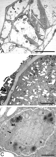
Table I. Summary of developmental variations in the anther abortive pattern of Consolea pistillate flowers at stage 2 (early prophase I).
Figure 4 A–F. TEM micrographs of Consolea picardae male‐sterile anther at Stage 2. A. Anther wall showing four layers: epidermis (e), endothecium (en), middle layer (ml) and a radially enlarged tapetum (tp). B. Microspore mother cell (mmc) at an early stage 2, before the tapetal cells start invading the locule, note the occasional vacuoles and the normal appearance of the cytoplasm and its contents. C. Tapetal cell showing peculiar rER configuration (arrow), and normal looking mitochondria (arrow head). D. mmc after tapetal cells begin radial elongation and compress the mmc; note the presence of numerous small vacuoles. E. Tapetal cell with almost circular rER configuration (arrow) encircling a portion of cytoplasm, a mitochondrium, and two Golgi. F. Tapetal cytoplasm with highly compressed and elongate rER cisternae (arrow) and normal looking mitochondria (arrow head). Scale bar – 2 µm (A, B, D); 1 µm (C); 0.5 µm (E, F).
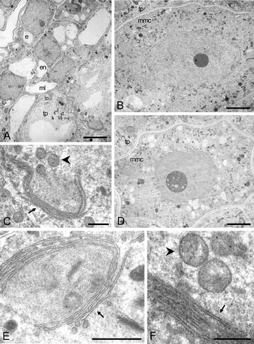
Figure 5 TEM micrograph montage ofConsolea picardae male‐sterile aborted anther. The anther wall is composed of epidermis (e), endothecium (en), middle layer (ml) and radially enlarged tapetum (tp). Both ml and tp cells are devoid of cytoplasm except for a thin layer adjacent to the cell wall; some lipid globules, rER remnants, and Golgi can be recognized. Scale bar – 2.5 µm.
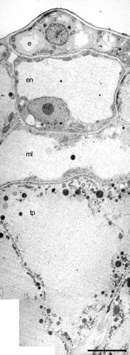
Figure 6 A–J. Bright field micrographs of Consolea millspaughii, C. moniliformis, C. nashii, C. rubescens and C. spinosissima male‐sterile anthers at stage 2 (left column) and at abortion (right column). A–B. C. millspaughii. A. Anther wall composed of epidermis (e), endothecium (en), middle layer (ml), and radially enlarged tapetum (tp); notice the single large vacuole in the tp cells. B. Remaining anther wall layers: e, en and radially enlarged ml; within the anther locule; some mmc and tp cell remmants are visible. C–D. C. moniliformis. C. e, en, ml and radially enlarged tp with single large vacuoles; the mmc's show some signs of degeneration. D. All anther layers except e, invading the locule, and in the center, a dark mass representing all that is left of the mmc's. E–F. C. nashii. E. e, en, ml, and radially enlarged tp with single large vacuoles. F. tp cells enlarged and invading the locule. G–H. C. rubescens. G. e, en, ml and somewhat enlarged tp cells with numerous small vacuoles. H. en and ml layers greatly enlarged and invading the locule; in the center, remnants of mmc's and tp cells are visible as dark masses. I–J. C. spinosissima. I. e, en, ml and somewhat radially enlarged tp with numerous small vacuoles. J. Enlarged en and ml invading the locule, and in the center, remnants of aborted tp cells and mmc's. Scale bars – 25 µm (A–J).
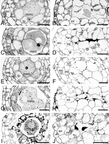
Figure 7 A–I. TEM micrographs of Consolea picardae, C. millspaughii, C. nashii, and C. rubescens male‐sterile anthers at Stage 2. A. Montage of C. picardae anther with a variant of the standard type of abortion; the middle layer (ml) and tapetal (tp) layers show a dense, dark cytoplasm in somewhat flattened cells while the epidermis (e) and endothecium (en) look healthy. B. C. millspaughii detail of microspore mother cell (mmc) nucleus and cytoplasm, a synaptonemal complex (arrow) is clearly visible inside the nucleus. C–F. C. rubescens. C. Detail of mmc nucleus with nucleolus; a synaptonemal complex (arrow) can be seen close to the nuclear membrane. D. Detail of tapetal cell with two nuclei. E–F. Detail of tapetal cell cytoplasm. E. rER with dilated cisternae that look almost like vescicles (solid arrow), also small vacuoles (v) and healthy looking mitochondria (arrowhead). F. Net‐like configuration of rER. G–H. C. nashii, tapetal cell cytoplasm. G. Clustered and elongated rER cisternae (solid arrow). H. Dilated rER cisternae (arrow). I. C. millspaughii tapetal cell with net‐like rER (arrow), abundant lipid globules (lp) and healthy looking mitochondria (arrowhead). Scale bar – 2 µm (A, D); 1 µm (C, I); 0.5 µm (B); 0.4 µm (E, H); 0.3 µm (G); 0.25 µm (F).
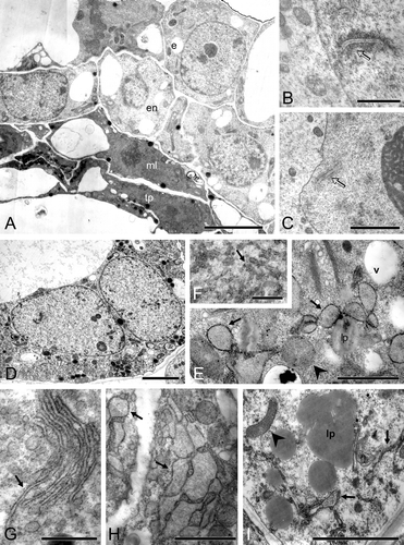
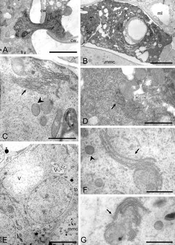
Figure 8 A–G. TEM micrographs of Consolea moniliformis and C. spinosissima male‐sterile anthers at Stage 2. A–D. C. moniliformis. A. Detail of a nucleus (n) traversing a cytoplasmic channel between two mmc's cell walls (cw). B–D. Tapetal cell. B. Somewhat enlarged tapetal cell (tp) with very dense cytoplasm and small vacuoles. C. Cytoplasm detail showing short compact rER cisternae forming dense stacks (solid arrow) and healthy looking mitochondria (arrowhead). D. Dense cytoplasm with some small vacuoles, some rER (solid arrow) and abundant free ribosomes. E–G. C. spinosissima tapetal cell (tp). E. Enlarged tp with vacuoles (v) and single nucleus. F. Cytoplasm detail showing elongated rER cisternae (solid arrow) encircling regions of cytoplasm, and healthy looking mitochondria (arrowhead). G. Cytoplasm detail showing nearly circular rER configuration (solid arrow). Scale bar – 2 µm (B, E); 1 µm (A); 0.5 µm (D, F, G); 0.4 µm (C).