Figures & data
Fig. 1. SEM micrographs of tapetum and pollen grains of Bromeliaceae. A – C. Pollen grains and tapetum with orbicules/lipid: (A) Orthophytum vagans, (B) Aechmea conglomerata, (C) Guzmania madisonii. D, E. Young pollen grains with orbicules/lipid: (D) Puya floccosa, (E) Aechmea conglomerata. F, G. Mature pollen grains: (F) Quesnelia edmundoi, (G) Dyckia hebdingii. O=possible orbicule. Bars– 15 μm (A, F, G); 25 μm (B, C); 5 μm (D, E).
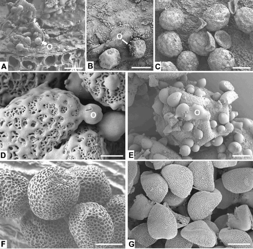
Fig. 2. Developmental stages of Bromeliaceae anthers in cross section (LM). A. Dyckia hebdingii, young anther possessing archesporial cells in the hypodermal tissue of each lobe. B. Tillandsia dura, young anthers, possessing parietal and primary sporogenous tissues. C. Dyckia hebdingii, with microsporocytes. D – F. Pitcairnia paniculata, development of the microsporocytes. AC=archesporial cells, En=endothecium, M=middle layer, MMC=microsporocyte, P=parietal cell layer, PS=primary sporogenous tissue, T=tapetum. Bars – 20 μm (A, E); 50 μm (B, C, D, F).
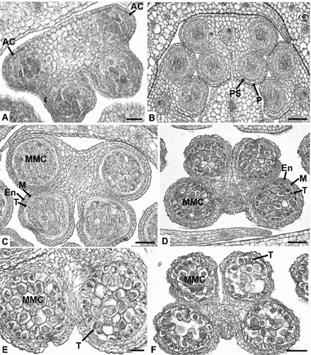
Fig. 3. Bromeliaceae anthers in cross section (LM). A. Puya floccosa, dyads and tetrads of microspores. B, C. Quesnelia edmundoi, enlarged tapetal cells and dyads and tetrads of microspores. D. Guzmania madisonii, tapetal material inside the locule. E. Pitcairnia paniculata, possible orbicules on the tapetal cells. F. Brocchinia reducta, possible orbicules on the tapetal cells. M=microspore, O=possible orbicule, T=tapetum, TM=tapetal material. Bars – 20 μm.
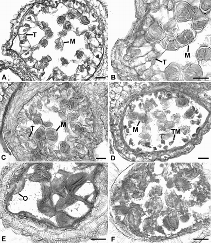
Fig. 4. Bromeliaceae anthers in cross section (LM). A. Orthophytum vagans, degenerating tapetal cells inside the locule. B. Aechmea conglomerata, possible orbicules on the microspores. C. Tillandsia dura, mature anthers. D. Brocchinia reducta, stamens in two whorls. E. Guzmania madisonii, introrse longitudinally dehiscent anthers. Anther walls consist of two cell layers (arrowed). Note papillate epidermis and presence of crystals. F. Brocchinia reducta, mature anthers consist of two layers, a spirally thickened endothecium and a papillate epidermis. C=crystals, En=endodermis, Ep=epidermis, M=microspore, O=possible orbicule, TM=tapetal material. Bars – 50 μm.
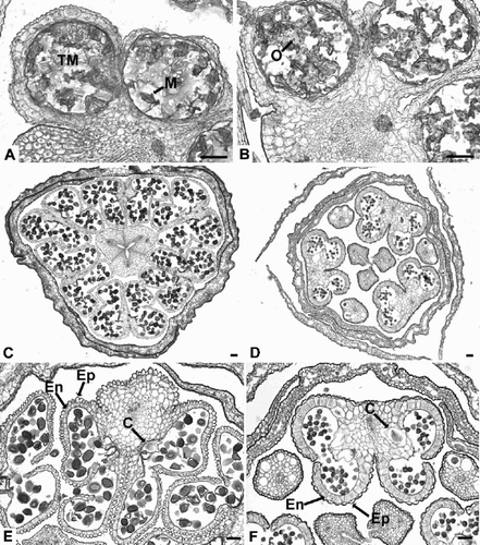
Fig. 5. Anthers of Bromeliaceae. A–C. Aechmea aquilega: (A) Anther locule with tetragonal tetrads surrounded by tapetal cells (LM); (B) Tapetal cell with a large nucleus adjacent to a tetrad (TEM); (C) Anther locule with sterile pollen (empty exines) surrounded by tapetal debris (LM). D – F. Aechmea abbreviata: (D) Anther locule with free microspores recently released from the callose wall and tapetal material inside the locule (LM); (E) Detail of microspores (TEM); (F) Remains of a tapetal cell at the edge of the anther locule (TEM). C=callose, M=microspore of tetrad, N=nucleus, T=tapetum, TM=tapetal material. Bars – 30 μm (A, C, D); 2 μm (B, E, F).
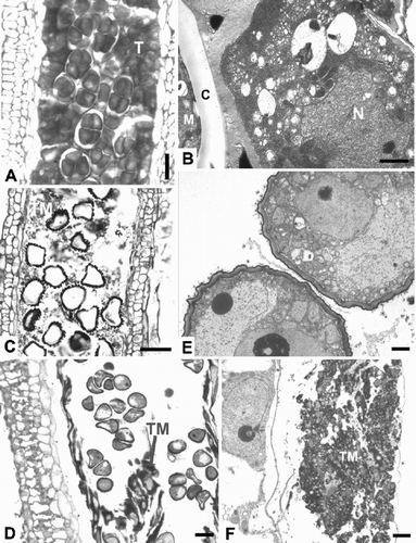
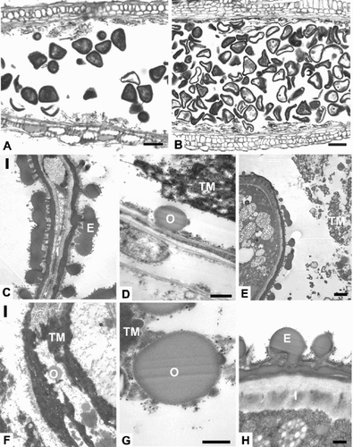
Fig. 6. Aechmea dichlamydea var. trinitensis. A. Anther locule containing pollen with the cytoplasm darkly stained (LM). B. Anther locule containing collapsed sterile pollen lacking cytoplasm (LM). C. Collapsed sterile pollen with exine and intine (TEM). D. A possible orbicule and remains of the tapetum adjacent to the locular wall (TEM). E. A mature pollen grain inside the locule with some tapetal material (TEM). F. Remains of the tapetum with possible orbicules (TEM). G. Detail of a possible orbicule (TEM). H. Detail of the pollen exine and intine (TEM). E=exine, I=intine, O=possible orbicule, TM=tapetal material. Bars – 30 μm (A, B); 1 μm (C, F); 0.5 μm (D, G, H); 2 μm (E).