Figures & data
Figure 1 Comparative equatorial diameters(µm) of spores.
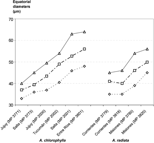
Figure 2 SEM micrographs ofAdiantopsis chlorophylla (Sw.) Fée and A. radiata (L.) Fée. A–D. A. chlorophylla. A. A spore in proximal view. The ornamentation is echinate. The laesurae are short and are evident on the perispore surface. B. A spore in distal view. The ornamentation is echinate. Many processes are fused. The elements of the ornamentation are densely distributed. C. Detail with a higher magnification of the echinate distal surface; the echinae are robust, their tips show varieties of shapes, while bases are wide and uniform. The echinae bases are complex and consist of rodlets fused that form a net (arrow). D. Detail of a fracture through the perispore (P). Below the perispore the exospore (E) is evident. It has a slightly rugulate surface. The two levels of the perispore are clearly distinguished: a basal level formed of a thin layer adhered to the exospore with rodlets, and a high level composed of echinae. E–H. A. radiata. E. A spore in proximal view. The ornamentation is echinate and the surface between echinae has a complex net of rodlets. The laesurae are short. F. A spore in distal view with ornamentation similar to the proximal face. G & H. Details of the distal surface at a higher magnification. G. The echinae are thin and acute, their bases are formed of radial rodlets fused to those of contiguous processes. H. In the basal level between the rodlets there are spherical elements of varied sizes. Scale bar – 10 µm (A, B, E, F, H); 1 µm (C, D, G).
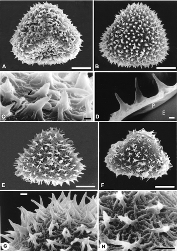
Figure 3 TEM ofAdiantopsis chlorophylla (Sw.) Fée. A–F. The exospore (E) is evident in A to E, its margin is smooth and in section its structure is homogenous. The exospore has uniform thickness and is less contrasted than the perispore. The perispore is two‐layered. The P1 layer is adhered to the exospore and forms the main part of this wall. The layer P2 is thin and covers the inner and outer surfaces of P1. A. In the middle part of layer P1 there are chambers (C) and radial elements (asterisk) of different thickness while this layer is compact towards the outside. The layer P2 is uniform in thickness and its margin is irregular. B. A section that shows spaced elements and those which form the bases of the complex processes of layer P1. Two processes (asterisks) in transversal section can be seen on the right. In both cases the layer P2 covers P1. Spaces of different width are evident in basal part of the processes (star). The spaces are covered with the P2 layer. In the contact between perispore and exospore, short superimposed plates are evident. C. This section crosses a zone with low elements, an interruption in the perispore is also evident that is continuous with a channel (arrowhead) in the exospore (E). This channel is larger in diameter than those at the inner part, and it is filled with helicoidal material of the same contrast as the perispore. (S) points spherules embodied between P1 and P2. D. Section at the laesura level, the exospore thickens and is continuous over the aperture. The perispore is also continuous over the aperture and its margin is irregular according to the sector sectioned. The perispore layer P2 covers P1 and is also continuous on the laesura. At the inner part of the laesura in the exospore, there are thin channels tangentially arranged (arrowheads); other channels are visible in the middle and inner part of the exospore with mainly radial orientation and apparently there exist others that connect each other. E. Detail of a section that shows the exospore and perispore. The layer P1 and P2 are clearly evident. There is a process with a chamber (C) in its base. The inner part of the chamber is covered by P2. F. An echina formed of P1 with a central region of low density (star). The process is covered by P2 which shows different degrees of development on the same spore. Scale bar – 1 µm (A–F).
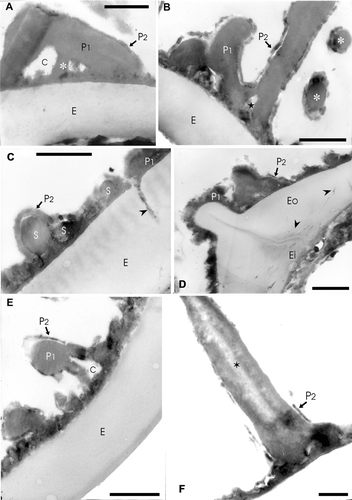
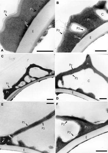
Figure 4 TEM ofA. radiata (L.) Fée A–F: In all the cases the exospore (E) margin is smooth and its structure is homogenous. The perispore is more contrasted than the exospore; it is composed of two layers: P1 and P2. P1 constitutes the main part of this wall. A. A scale (Sc) is evident in the inner part of P1. The layer P2 is very thin and covers P1. B. In this section chambers (C) within the layer P1 can be seen, they are surrounded by a less contrasted layer. A scale (Sc) is evident in the inner part of P1. C. In this case chambers (C) can be seen within P1. The (P2) layer is thin, less contrasted than layer P1 and only visible in some places. D. Spine with a basal chamber (C) in detail. The layer P2 covers the inner and outer surfaces of P1. E. The section shows a process of high length formed mainly by P1. A scale (Sc) is evident in the inner part of P1, near the left limit. F. In this section there are two perispore processes of different height, they are formed by P1 and covered by P2. A dark extensive lamina is visible between the exospore and perispore. Scale bar – 1 µm (A–E).