Figures & data
Figure 1 Geological scheme of the Mesozoic outcrops in the Fuentelsaz area(FZ) and the location of the sites mentioned in the text.
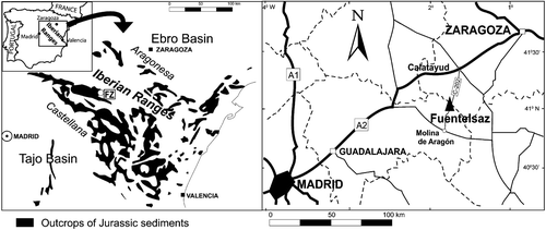
Figure 2 Stratigraphic succession of the Fuentelsaz section and the relative distribution of theClassopollis species.
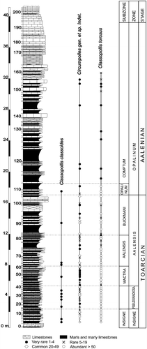
Figure 3 A–H. Type A: Classopollis classoides. (A) Light microscope (LM) photomicrograph of an individual pollen grain of C. classoides in polar view; (B) Confocal scanning laser microscope (CSLM) photomicrograph of the same grain obtained by maximum projection of the optical slice stack along the vertical axis. Note that the white spots (black arrow) correspond to sexine elements; (C) LM photomicrograph of another specimen of C. classoides in equatorial view; (D & E). CSLM partial maximum projection of sections of the same grain as in (C). Each composite view represents a 0.8 µm‐thick section of the grain. Note the anastomosing system (black arrow) formed by the sexine elements; (F) CSLM full maximum projection of the same grain shown in (C). Note the well defined rimula and equatorial endostriae; (G) CSLM detail of the same grain as in (C). Note the thinning of the sexine at the level of the rimula (arrow). (H) Three‐dimensional (3‐D) rendering based on CSLM sections of the same grain as in (C). Note the psilate nature of the sculpture and the emerging lips of the trilete scar.
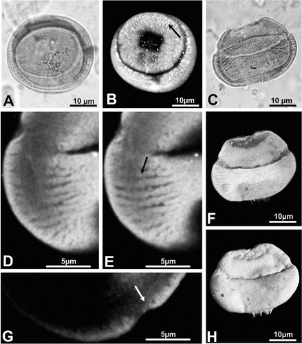
Figure 4 A, B & D, E. Type B: Classopollis torosus. C. Type C: Circumpolles gen. et sp. indet. (R – rimula; T – trilete scar; P – distal pore). (A) LM photomicrograph of a tetrad of pollen grains attributed to C. torosus; (B) LM photomicrograph of a single pollen grain of C. torosus; (C) LM photomicrograph of a tetrad of pollen grains attributed to Circumpolles gen. et sp. indet.; (D) 3‐D rendering based on CSLM sections of the same grain as in (B). Note the scabrate nature of the sculpture; (E) 3‐D rendering based on CSLM sections of the same grain as in (B) but showing its back side. Note the pore and trilete scar.
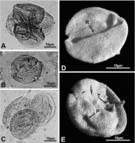
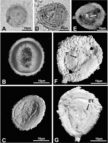
Figure 5 A–G. Type C: Circumpolles gen. et sp. indet. (R – rimula; T – trilete scar; ET – thickened annulus). (A) LM photomicrograph of an individual pollen grain attributed to Circumpolles gen. et sp. indet.; (B) CSLM maximum vertical projection of sections of the same grain; (C) 3‐D rendering based on CSLM sections of the same grain showing the proximal side; (D) LM photomicrograph of another specimen of Circumpolles gen. et sp. indet.; (E) CSLM maximum vertical projection of sections of the same grain; (F) 3‐D rendering based on CSLM sections of the same grain viewed from its proximal side. The triangular area represents the trilete scar; (G) 3‐D rendering based on CSLM sections of the same grain viewed from its distal side. The pore has a thickened annulus.