Figures & data
Figure 2. Clockhousea capelensis. SEM. A. Distal surface of specimen, sample DJBCl88/10, preparation MCM193, BGS reg. MPK13806. B. Lateral view of spore, part of narrow triradiate ridge visible (arrow), DJBCl88/10, MCM193, MPK13807. C. Specimen with pronounced triradiate ridge that shows evidence of damage in the form of abrasion of sculptural elements, especially towards proximal pole, DJBCl88/11, MFP166, MPK13808. D. Well-preserved specimen sculptured predominantly by closely spaced verrucae, but with scattered composite baculate elements mainly on proximal surface, especially towards triradiate ridge, DJBCl88/11, MFP166, MPK13809. E. Close-up of proximal polar area of spore depicted in C. F. Example of a spore sculptured solely by closely spaced verrucae and assumed to be an atypical, immature or aberrant form of the species, DJBCl88/10, MCM193, MPK13810. Scale bars – 100 μm (A–D, F); 10 μm (E).
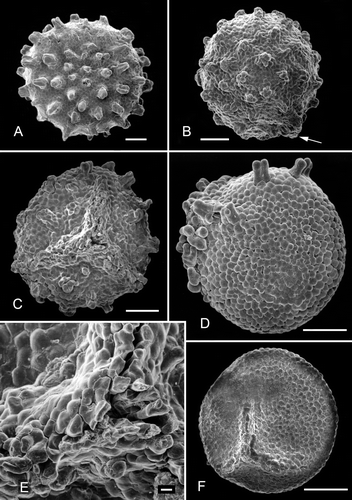
Figure 3. Clockhousea capelensis. SEM. A. Detail of spore depicted in showing ‘negative reticulum’ and composite baculate process, sample DJBCl88/10, preparation MCM193, BGS reg. MPK13811. B–F. Details of specimen broken in half, DJBCl88/10, MCM193, MPK13812: B. Components of ‘negative reticulum’ become narrower towards interior surface of wall, giving them a clavate appearance; they are attached to a thin inner, perforated layer; C. Detail of part of inner surface of perforated layer; D. General view of part of broken specimen; E. Perforations of another part of inner layer viewed more obliquely than in C; F. Part of wall showing an exinal extension that consists of single baculate and spinose elements that are not fused together. Scale bars – 10 μm.
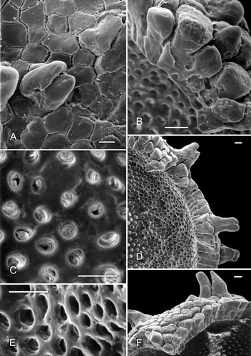
Figure 1. Clockhousea capelensis gen. et sp. nov. LM. A, B. Holotype, sample DJBCl88/10, preparation MCM193, BGS reg. MPK13804, photographed under reflected light: A. Proximal view, triradiate ridge unclear owing to the profusion of bacula, which mask it; B. Distal surface; C, D. A second, smaller specimen, DJBCl88/10, MCM193, MPK13805: C. Proximal view, triradiate ridge faintly discernible; D. Distal surface. Scale bar – 100 μm.
Figure 4. A–D, F. Clockhousea capelensis. SEM. A–C. Broken specimen, sample DJBCl88/10, preparation MCM193, BGS reg. MPK13813: A. Close-up of inner surface of exine in vicinity of triradiate suture, and possibly also a thin layer beneath it within suture (arrow); B. Detail of inner suface of exine; unlike the perforations shown in , E, these are less clearly delineated because the surface is much more uneven; C. General view of whole specimen. D. Detail of top of clavate element of exine that has suffered from extensive microbial degradation, same specimen as that in . F. Cross-section of spore that has been broken in half, showing a complex of baculate and other exinal elements bordering triradiate suture and a more or less smooth inner surface of what appears to be a thin inner layer of exine, DJBCl88/10, MCM193, MPK13814. E. An example of what may or may not be an ‘inner body’ (the inner separable layer) of a specimen of C. capelensis that has lost its outer wall, DJBCl88/10, MCM193, MPK13815. Scale bars – 100 μm (A, C, E, F); 10 μm (B); 1 μm (D).
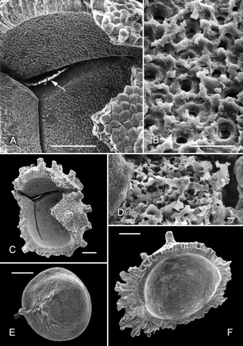
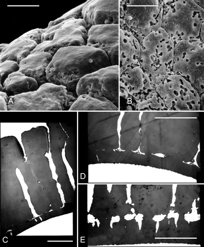
Figure 5. A, B. Scanning electron micrographs of parts of the ‘negatively reticulate’ surface of two examples of Clockhousea capelensis showing the beginnings of, and more serious, microbial degradation respectively: A. Same specimen as that in B. Sample DJBCl88/10, preparation MCM193, BGS reg. MPK13816. C–E. Transmission electron micrographs of parts of the exine of two specimens of C. capelensis showing its structure. The columnar elements become much narrower just above their points of attachment to the inner perforated layer. The specimens were difficult to cut and none of the sections passes through the centre of a perforation: C, D. Cross-sections of same specimen; E. Slightly oblique cross-section of a second specimen. Scale bars – 10 μm.