Figures & data
Figure 1. Locality map. A. Location of Ethiopia in Africa. B. The study area, Gonder, and the current capital city, Addis Ababa. Major rivers are shown, including the Blue Nile and its source, Lake Tana, to the immediate south of the study area. C. Location of the Guang and Hauga Rivers, and measured section shown in .
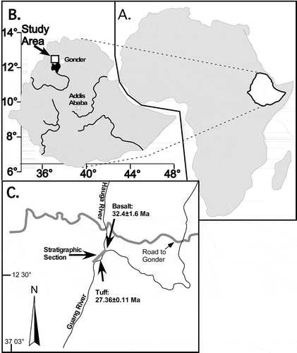
Figure 2. Stratigraphic section, measured along the Guang River. Stars indicate stratum that produced the Sclerosperma fossil pollen, and the fossil leaf locality, CH41, is labelled and marked by a leaf icon. Radioisotopic dates are shown near the base and top of the section.
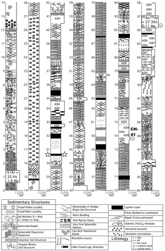
Table I. Pollen morphology of extant and fossil Sclerosperma.
Figure 3. Light microscopy (LM) (A) and scanning electron microscopy (SEM) (B–I) micrographs of Sclerosperma protomannii sp. nov. (holotype: IPUW 7513/223). A. Pollen grain in polar view (upper in high focus and middle in optical cross-section) and equatorial view (lower). B. Pollen grain in polar view, distal side. C. Pollen grain in polar view, proximal side. D. Close-up of apex with aperture, distal side. E. Close-up of apex, proximal side. F. Close-up of central polar area, distal side. G. Close-up of central polar area, proximal side. H. Close-up of interapertural area, distal side. G. Close-up of interapertural area, proximal side. Scale bars – 10 µm (A–C), 1 µm (D–I).
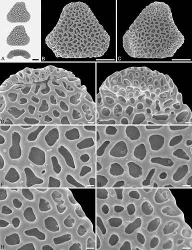
Figure 4. Light microscopy (LM) (A) and scanning electron microscopy (SEM) (B–E) micrographs of Sclerosperma protomannii sp. nov. (paratype: IPUW 7513/224). A. Pollen grain in polar view (optical cross-section). B. Pollen grain in polar view, distal side. C. Pollen grain in polar view, proximal side. D. Close-up of central polar area, distal side. E. Close-up of interapertural area, proximal side. LM (F) and SEM (G–J) micrographs of S. protomannii sp. nov. (paratype: IPUW 7513/225). F. Pollen grain in polar view (high focus). G. Pollen grain in polar view, distal side. H. Pollen grain in polar view, proximal side. I. Close-up of apex with aperture, distal side. J. Close-up of apex, proximal side. Scale bars – 10 µm (A–C, F–H), 1 µm (D, E, I, J).
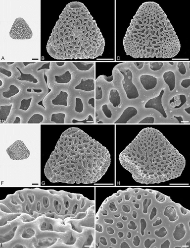
Figure 5. Light microscopy (LM) (A) and scanning electron microscopy (SEM) (B–E) micrographs of Sclerosperma protomannii sp. nov. (paratype: IPUW 7513/226). A. Pollen grain in polar view (upper, high focus) and equatorial view (lower). B. Pollen grain in polar view, distal side. C. Pollen grain in polar view, proximal side. D. Close-up of apex with aperture, distal side. E. Close-up of central polar area, distal side. LM (F) and SEM (G–J) micrographs of S. protoprofizianum sp. nov. (holotype: IPUW 7513/227). F. Pollen grain in polar view (high focus). G. Pollen grain in polar view, distal side. H. Pollen grain in polar view, proximal side. I. Close-up of apex with aperture, distal side. J. Close-up of central polar area, distal side. Scale bars – 10 µm (A–C, F–H), 1 µm (D, E, I, J).
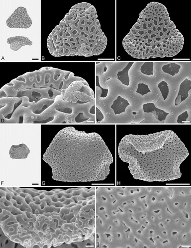
Table II. Fossil record of Sclerosperma
Figure 6. Light microscopy (LM) (A) and scanning electron microscopy (SEM) (B–E) micrographs of Sclerosperma protoprofizianum sp. nov. (paratype: IPUW 7513/228). A. Pollen grain in polar view (high focus). B. Pollen grain in polar view, distal side. C. Pollen grain in polar view, proximal side. D. Close-up of apex with aperture, distal side. E. Close-up of central polar area, distal side. LM (F) and SEM (G–J) micrographs of S. protoprofizianum sp. nov. (paratype: IPUW 7513/229). F. Pollen grain in polar view (high focus). G. Pollen grain in polar view, distal side. H. Pollen grain in polar view, proximal side. I. Close-up of apex, proximal side. J. Close-up of central polar area, distal side. Scale bars – 10 µm (A–C, F–H), 1 µm (D, E, I, J).
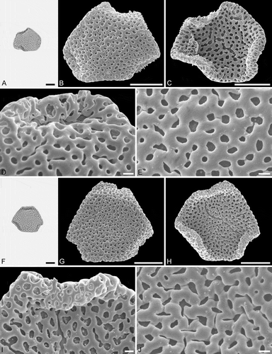
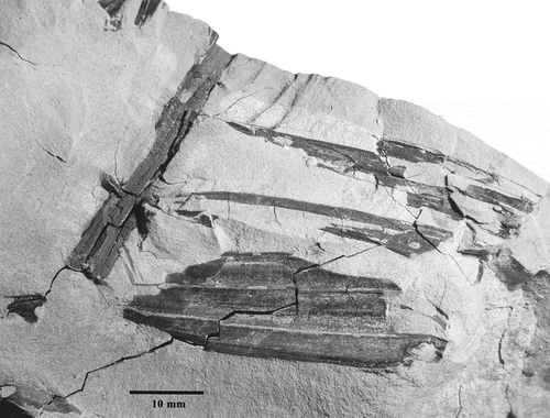
Figure 7. Fossil leaf fragment from locality CH41, specimen 9A (CH41-9A), collected at 74 m above the base of the measured Guang River section ().