Figures & data
Table I. Centauropsis dimensions (μm) pollen grains in equatorial and polar view using light microscopy.
Table II. Dimensions (μm) pollen grains aperture, spine, reticulum and exine layers in equatorial view using light microscopy.
Figure 1. Light microscopy of Centauropsis Bojer ex DC. pollen grains. A‒C. Centauropsis antanossi (Scott Elliot) Humbert. A. Detail of the colporus, endoaperture and surface, equatorial view. B. Apertures 3-colporate, long and acute apices and apocolpium surface. C. Optical section, equatorial view. D‒F. Centauropsis cuspidata Humbert. D. Detail of the colporus, endoaperture and surface, equatorial view. E. Apertures 3-colporate, long and acute apices and apocolpium surface. F. Optical section, equatorial view. G‒H. Centauropsis decaryi Humbert. G. Detail of the colporus, endoaperture and surface, equatorial view. H. Apertures 3-colporate, long and acute apices and apocolpium surface. I. Optical section, equatorial view. J‒L. Centauropsis fruticosa Bojer ex DC. J. Detail of the colporus, endoaperture and surface, equatorial view. K. Apertures 3-colporate, long and acute apices and apocolpium surface. L. Optical section, equatorial view. Scale bars – 10 μm (A‒C, G‒L), 8 μm (D‒F).
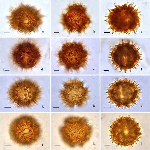
Figure 2. Scanning electron microscopy of Centauropsis Bojer ex DC. pollen grains. A‒C. Centauropsis antanossi (Scott Elliot) Humbert. A. General view of the mesocolpium, equatorial view. B. Detail of apertural membrane of the colporus, margo, and mesocolpium. C. Fractured pollen grain, structure of exine layers. D‒F. Centauropsis cuspidata Humbert. D. General view of the apertural area and mesocolpium, equatorial view. E. Detail of apocolpium, polar view. F. Detail of the spines and lophae on mesocolpium, equatorial view. G–I. Centauropsis decaryi Humbert. G. General view of the mesocolpium, equatorial view. H. General view of the apocolpium, polar view. I. Fractured pollen grain, structure of exine layers. J‒L. Centauropsis fruticosa Bojer ex DC. J. General view of the apertural area and mesocolpium, equatorial view. K. Detail of apocolpium, polar view L. Detail of apertural membrane of the colporus, margo, and mesocolpium. Scale bars – 10 μm (A, D, G, H, J), 1 μm (B, C, E, F, I, K, L).
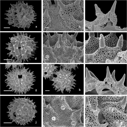
Figure 3. Light microscopy of Centauropsis Bojer ex DC. pollen grains. A‒C. Centauropsis fruticosa var. baronii Humbert. A. Detail of the colporus, endoaperture and surface, equatorial view. B. Apertures 3-colporate, long and acute apices and apocolpium surface. C. Optical section, equatorial view. D‒F. Centauropsis perrieri Humbert. D. Detail of the colporus, endoaperture and surface, equatorial view. E. Apertures 3-colporate, long and acute apices and apocolpium surface. F. Optical section, equatorial view. G‒I. Centauropsis rhaponticoides Drake. G. Detail of the colporus, endoaperture and surface, equatorial view. H. Apertures 3-colporate, long and acute apices and apocolpium surface. I. Optical section, equatorial view. Scale bars – 10 μm (A‒C), 8 μm (D‒I).
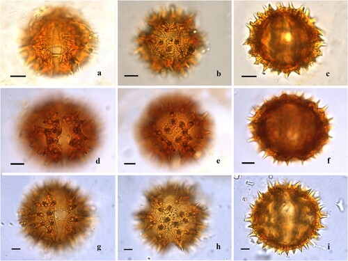
Figure 4. Scanning electron microscopy of Centauropsis Bojer ex DC. pollen grains. A‒C. Centauropsis fruticosa var. baronii Humbert. A. General view of the apertural area, mesocolpium, and apocolpium, inclined equatorial view. B. Detail of the spines, lophae and microspines on mesocolpium, equatorial view. C. Fractured pollen grain, structure of exine layers. D‒F. Centauropsis perrieri Humbert. D. General view of the apertural area, mesocolpium, equatorial view. E. Detail of the apocolpium, polar view. F. Detail of apertural membrane of the colporus, margo, and mesocolpium. G‒I. Centauropsis rhaponticoides Drake. G. General view of the apertural area, mesocolpium, equatorial view. H. Detail of the apocolpium, polar view. I. Detail of the spines on mesocolpium, equatorial view. J, K. Fractured pollen grain, structure of exine layers of Centauropsis fruticosa var. baronii Humbert. L. Fractured pollen grain, structure of spine of Centauropsis perrieri Humbert. Scale bars ‒ 10 μm (A, D, G), 1 μm (B, C, E, F, H‒L).
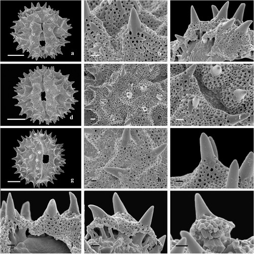
Table III. Morphology and ultrasculpture of Centauropsis pollen grains using light and scanning electron microscopy.
Figure 5. Principal component analysis biplot of the pollen grain metric variables and classes/indices of Centauropsis specimens.
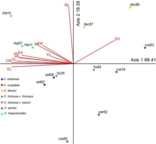
Table IV. Pearson and Kendall coefficients of pollen grain metric variables and classes/indices from the first two ordination axes of the principal component analysis (PCA) of Centoropsis species.
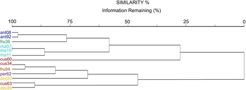
Figure 6. Dendrogram built from cluster analysis (Euclidean distance) for Centauropsis specimens, information remaining (%).