Figures & data
Table 1. Characteristics of liposomes
Table 2. Plasma coagulation activity as measured by PT and APTT
Figure 1 Activation of kallikrein-kinin cascade by HbVs. HbVs or saline were mixed with plasma as indicated ratio (v/v) at 37°C for 24 h. Appearance of digested S-HMWK was detected using western blot analysis as a result of kallikrein activation. Arrows indicate digested S-HMWK. A typical result of three independent assays is shown. H, S-HMWK; dH, digested S-HMWK; lanes 1, 5 and 9, saline; lanes 2, 6 and 10, DPPG-HbV; Lanes 3, 7 and 11, PEG-DPPG-HbV; lanes 4, 8 and 12, PEG-DPEA-HbV.
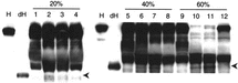
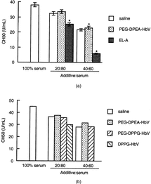
Figure 2 Consumption of complement by HbVs. HbVs, saline or EL-A were mixed with serum as indicated ratio (v/v) at 37°C for 24 h. A: Data are represented as the mean ± SEM using serum from five individuals. *Significantly different from saline (p < 0.05). B: Representative data are shown using one serum.