
Figures & data
Figure 1. (a) Computed tomography (CT) performed before intravenous thrombolysis (IVT) shows old lacunar infarctions bilaterally in the area under the cortex of the head. (b) Head CT shows low-density foci in the right frontal and temporal lobes with minor hemorrhaging (no space-occupying effect) on the second day after IVT. (c) Chest CT shows inward displacement of the calcified plaques in the thoracic aortic arch (arrow) and suspicious double lumens from the aortic arch to the descending aorta, suggesting aortic dissection (AD). (d) CT angiography of the thoracoabdominal aorta shows a thoracoabdominal AD (DeBakey type I), with the AD extending from the aortic root to the brachiocephalic trunk (e) and then to the right common carotid artery. (f) Intimal slices split the lumen into two cavities in the aortic arch.
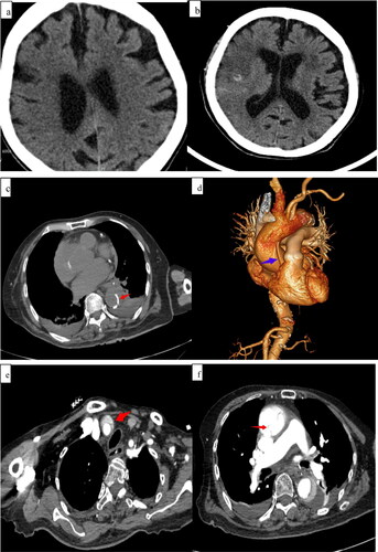
Table 1. Coagulation function before and after intravenous thrombolysis.
Table 2. Details of cases that underwent IVT with rt-PA for AD-related acute ischemic stroke.