
Figures & data
Figure 1. Patient 1. (a) Emergency head computed tomography before intravenous thrombolysis showing no obvious abnormality. (b) Brain magnetic resonance angiography (MRA) showing no obvious abnormality. (c) Contrast-enhanced transcranial Doppler (c-TCD) showing a signal of a microbubble that overflowed in the resting state. (d) Contrast-enhanced transthoracic echocardiography showing 20–30 microbubbles per section in the resting state in the left atrium at the second cardiac cycle. Patient 2. (e) Diffusion-weighted imaging sequence showing an acute infarction in the left temporal lobe and frontotemporal junction. (f) MRA showing severe stenosis of the M2 segment of the left middle cerebral artery (MCA), which had sparse distal branches. (g) c-TCD showing a small amount of microbubbles in the resting state. (h) Contrast-enhanced transesophageal ultrasound showing approximately 20 microbubbles per section per frame.
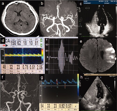
Figure 2. Sketch maps for different inspection effects of contrast-enhanced transcranial Doppler, transesophageal ultrasound, and transthoracic echocardiography (c-TCD, c-TEE and c-TTE, respectively). The middle cerebral artery detected by c-TCD is only a small branch of the aorta, and c-TTE/c-TEE may reveal more right-to-left shunts (more microbubbles) in the left atrium.
