
Figures & data
Figure 1. (a–h). Angiography showing acute basilar artery occlusion.
(a, b) The left vertebral arteriogram in the frontal and lateral view shows occlusion of the basilar artery, with a small amount of PICA flow compensating for the occluded segment of the basilar artery and intermittent visualization of the lumen of the basilar artery. (c, d) A frontal and lateral view of the right vertebral artery angiogram shows occlusion of the basilar artery with a small amount of antegrade compensation of the PICA branch. (e, f) A right internal carotid artery system frontal and lateral angiogram shows no significant abnormalities, and an embryonic posterior cerebral artery was observed. (g, h) A front and side view of the left internal carotid artery system showed no significant abnormalities, and the embryonic posterior cerebral artery was observed ().
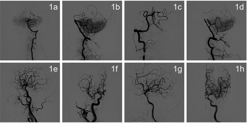
Figure 2. DSA imaging during drug-coated balloon dilatation angioplasty.
(a) A 5F*115 cm Navien intermediate catheter selected to the V2 end of the left vertebral artery for fixation. (b) A 3-m Synchro microguide with Rebar18 microcatheter fed through the occluded segment followed by intracatheter angiogram showing that the catheter was located in the true lumen of the vessel. (c) Ten minutes after 2.5 × 15 mm Gateway balloon dilation, the lumen of the stenosis was retracted significantly. (d) The microguide wire was fixed in the left superior cerebellar artery, and a 2.5 × 20 mm paclitaxel drug-coated balloon was used for dilation. (e, f) After drug-coated balloon dilation, the patient was observed for 50 min and re-imaged. The patient had good lumen formation of the basilar artery, no retraction, no artery dissection, distal flow patency, and mTICI grade 3.
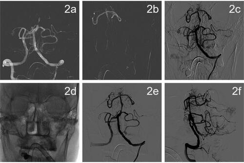
Figure 3. Cranial magnetic resonance imaging on postoperative day 3.
(a–c) Postoperative day 3 MRI DWI showed multiple fresh cerebral infarcts in the pons, cerebellar vermis, bilateral cerebellar hemispheres, frontoparietal temporal lobe, and corpus callosum. (d, e) MRA on postoperative day 3 showed local luminal stenosis of the middle basilar artery (shown by red arrows).
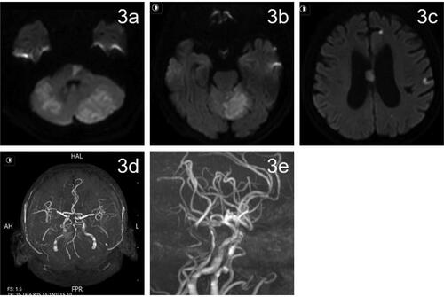
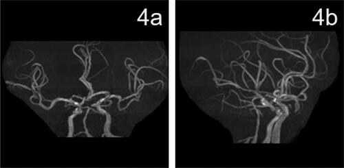
Figure 4. Head MRA results 3 months after surgery.
(a, b) MRA showed the basilar artery lumen was patent.
Data availability statement
All datasets presented in this study are included in the article.