113,174
Views
37
CrossRef citations to date
0
Altmetric
Articles

A new genus and species of Nematalycidae (Acari: Endeostigmata)

, , &
Pages 1359-1373 | Received 23 Apr 2013, Accepted 12 Sep 2013, Published online: 20 Feb 2014

Figures & data

Figure 1. Osperalycus tenerphagus sp. nov. Female: (A) dorsal view; (B) ventral view; (C) metapodosoma and genital region (D) genitalia – internal.
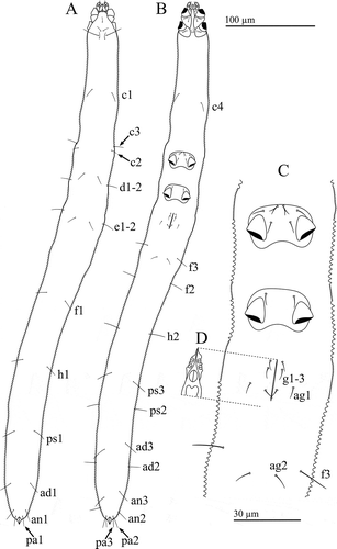
Figure 1. Osperalycus tenerphagus sp. nov. Female: (A) dorsal view; (B) ventral view; (C) metapodosoma and genital region (D) genitalia – internal.

Figure 2. Osperalycus tenerphagus sp. nov. Female, proterosoma: (A) dorsal view (chelicerae slanted downwards into the vessel, making them appear slightly shorter; broad bases of rutella partly behind chelicerae and vessel opening – shaded pale grey); (B) ventral view.
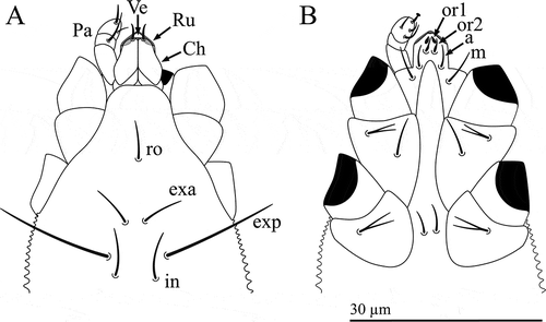
Figure 2. Osperalycus tenerphagus sp. nov. Female, proterosoma: (A) dorsal view (chelicerae slanted downwards into the vessel, making them appear slightly shorter; broad bases of rutella partly behind chelicerae and vessel opening – shaded pale grey); (B) ventral view.

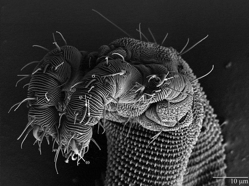
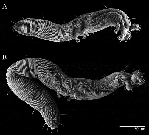
Figure 3. Osperalycus tenerphagus sp. nov. Integument: (A) extended, vertical view (showing palettes as thin and flat); (B) contracted, vertical view (showing interlocking palettes); (C) extended, diagonal view; (D) contracted, diagonal view.
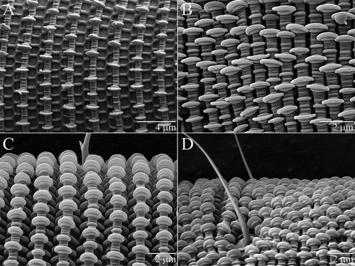
Figure 3. Osperalycus tenerphagus sp. nov. Integument: (A) extended, vertical view (showing palettes as thin and flat); (B) contracted, vertical view (showing interlocking palettes); (C) extended, diagonal view; (D) contracted, diagonal view.

Figure 4. Osperalycus tenerphagus sp. nov. Tritonymph, opisthosoma: (A) anal valves and ventral furrow, ventrolateral view; (B) anal valves, dorsal view; (C) anal valves, posterior view; (D) ventral furrow, close-up.
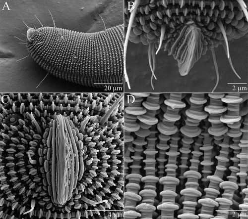
Figure 4. Osperalycus tenerphagus sp. nov. Tritonymph, opisthosoma: (A) anal valves and ventral furrow, ventrolateral view; (B) anal valves, dorsal view; (C) anal valves, posterior view; (D) ventral furrow, close-up.

Figure 5. Osperalycus tenerphagus sp. nov. Female: (A–E) tarsi I–IV; (A) tarsus I; (B) tarsus II, ε obscured; (C) tarsus II, ε visible; (D) tarsus III; (E) tarsus IV. Deutonymph: (F) tibia I. 
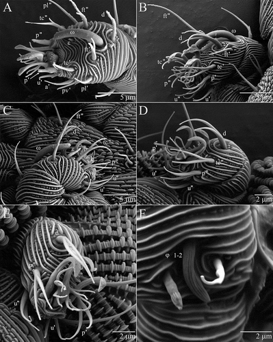
Figure 5. Osperalycus tenerphagus sp. nov. Female: (A–E) tarsi I–IV; (A) tarsus I; (B) tarsus II, ε obscured; (C) tarsus II, ε visible; (D) tarsus III; (E) tarsus IV. Deutonymph: (F) tibia I. 

Figure 6. Osperalycus tenerphagus sp. nov. Female: solenidia on legs I and II, female.

Figure 6. Osperalycus tenerphagus sp. nov. Female: solenidia on legs I and II, female.

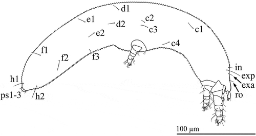
Figure 7. Osperalycus tenerphagus sp. nov. Larva: lateral view.

Figure 7. Osperalycus tenerphagus sp. nov. Larva: lateral view.

Figure 8. Osperalycus tenerphagus sp. nov. Nymphs: lateral view: (A) protonymph; (B) tritonymph.

Figure 8. Osperalycus tenerphagus sp. nov. Nymphs: lateral view: (A) protonymph; (B) tritonymph.

Table 1. Osperalycus tenerphagus sp. nov. Mean length of appendages and idiosoma across instars (µm).

Table 2. Osperalycus tenerphagus sp. nov. Distinguishing attributes of instars.

Table 3. Osperalycus tenerphagus sp. nov. Setal addition pattern for legs (including coxal fields) across instars.