Figures & data
Table 1. Data set of the octocorals collected from the El Pelado Equatorial Front (October 2018).
Figure 1. (a–c) Map of the sampling stations. (CENAIM-ESPOL, Bajo 40, Laberinto, Acuario, La Pared). Map credits: Divar Castro – CENAIM. (d) In situ rocky reefs of El Pelado which these species inhabit in sympatry. Exsitu Photos by D.C. Vergara and Juan A. Sánchez. Insitu Photos by Rubén Abad and Karla. B. Jaramillo.
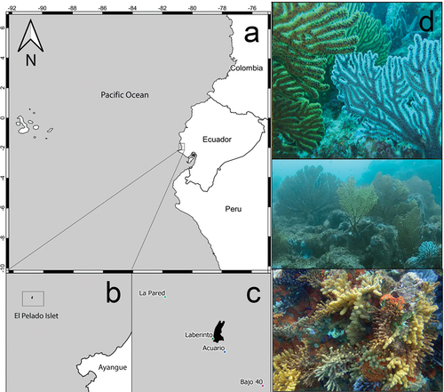
Figure 2. Underwater in situ record of Muricea hebes colony with (a) the white extended polyps, and (b) M. hebes with the polyps contracted. (c) Ex situ dry colony of M. hebes. (d) Location of colonies inside a cavern at the Bajo 40 site. (e) Close-up of the small tubular calyxes in a branch. Exsitu Photos by D.C. Vergara and Juan A. Sánchez. Insitu Photos by Rubén Abad and Karla. B. Jaramillo.
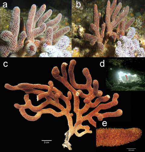
Figure 3. Underwater in situ record of Muricea echinata: (a) full colony with extended white polyps and close-up of the polyps at La Pared site. (b) Ex situ dry colony of M. echinata. (c) Close-up of the calyxes in a bifurcated branch. Exsitu Photos by D.C. Vergara and Juan A. Sánchez. Insitu Photos by Rubén Abad and Karla. B. Jaramillo.
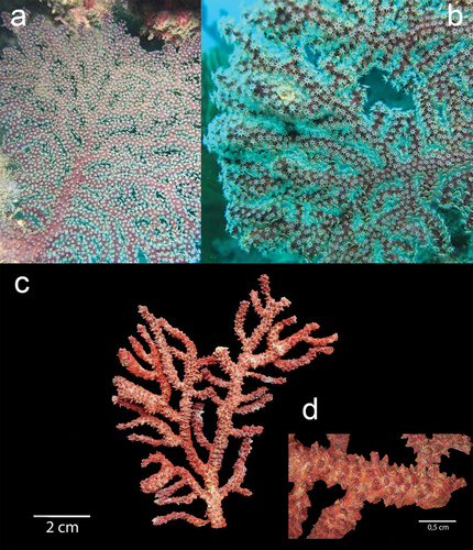
Figure 4. Underwater in situ record of Muricea robusta: (a) full colony with extended yellow polyps and close-up of the polyps in Bajo 40 site; and (b) with the polyps inside the colony. (c) Ex situ dry colony of M. robusta. (d) Close-up of the large orange calyxes in a branch. Exsitu Photos by D.C. Vergara and Juan A. Sánchez. Insitu Photos by Rubén Abad and Karla. B. Jaramillo.
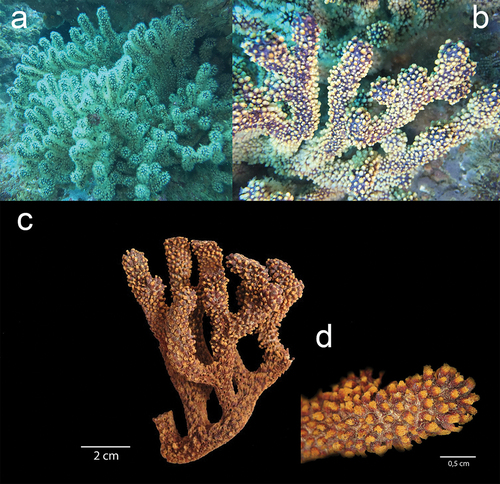
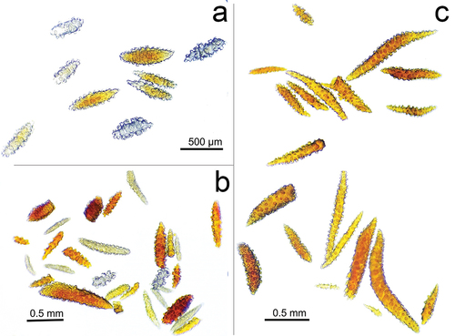
Figure 5. Sclerites of the three species (a) Muricea hebes, (b) Muricea robusta and (c) Muricea echinata collected at El Pelado Islet (REMAPE area). Sclerite Photos by Rubén Abad and Karla. B. Jaramillo.