Figures & data
Figure 1. Habitus of Spinozodium denisi (a, b) and S. khatlonicum sp. nov. (c, d), dorsal view. (a, c) females; (b, d) males. Scale bars = 0.5 mm.
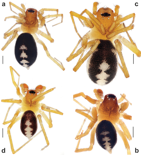
Figure 2. Male palps of Spinozodium denisi (a–c) and S. khatlonicum sp. nov. (d–f). (a, d) ventral view; (b, e) prolateral view; (c, f) dorsal view. Scale bars = 0.2 mm.
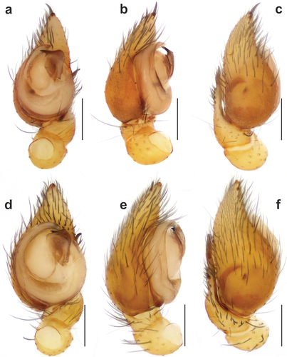
Figure 3. Scanning electron microscopy images of the male palps of Spinozodium khatlonicum sp. nov. (a–c) and S. denisi (d–g). (a, e) detail of embolus and tegular apophysis, anteroventral view; (b, d) ventral view; (c, f) retrolateral view; (g) cymbial trichobothrium. Scale bars = 0.1 mm, unless otherwise stated. Abbreviations: Cf – cymbial fold, Co – conductor, Em – embolus, Ep – process of embolus tip, Os – opening of the sperm duct, Ta – tegular apophysis, Tp – process of tegular apophysis, Tr – retrolateral arm of tegular apophysis, Tt – teeth of tegular apophysis.
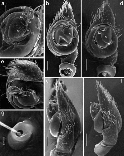
Figure 4. Spinozodium denisi (a, b) and S. khatlonicum sp. nov. (c, d). (a, c) male palp, retrolateral view; (b, d) intact epigyne, ventral view. Scale bars = 0.2 mm.
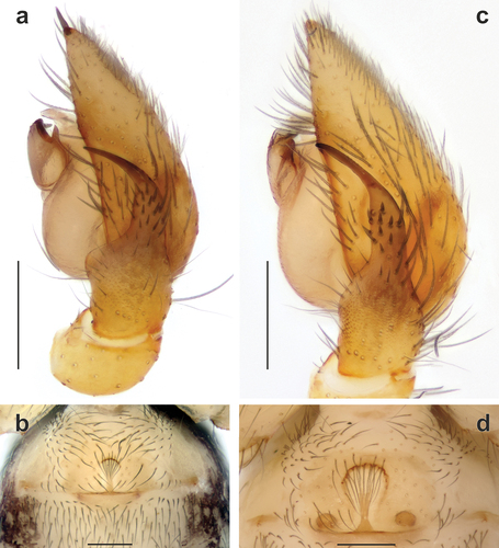
Figure 5. Macerated epigynes of Spinozodium denisi (a, b) and S. khatlonicum sp. nov. (c, d). (a, c) dorsal view; (b, d) ventral view. Scale bars = 0.2 mm. Abbreviations: Ah – anterior hood, Rh – head of receptacle, Sb – septal base, Ss – septal stem.
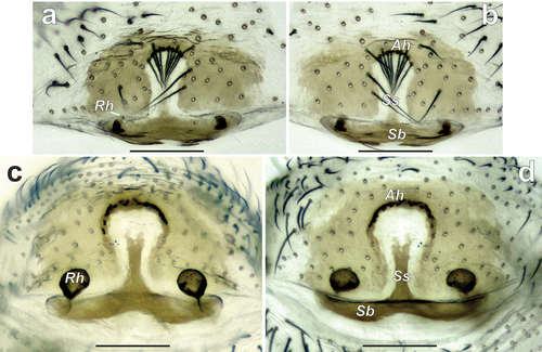
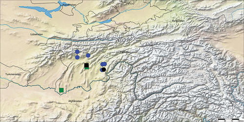
Figure 6. Collection localities of Spinozodium denisi (circles) and S. khatlonicum sp. nov. (squares). Black symbols indicate the type localities.