Figures & data
Figure 1. Anatomical position and constitution of the Phantom.
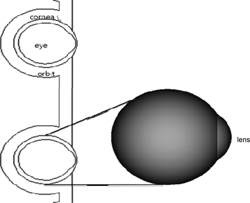
Table 1. Composition of the tissues for the phantoms.
Table 2. Estimation of the absorbed dose of the head, eyes, corneas, and lenses according to energy level.
Table 3. Estimation of the absorbed dose of the head, eyes, corneas, and lenses when using an eye shield (thickness of shield: 0.5 mm).
Table 4. Estimation of the absorbed dose of the head, eyes, corneas, and lenses when using an eye shield (thickness of shield: 1.0 mm).
Table 5. Estimation of the absorbed dose of the head, eyes, corneas, and lenses when using an eye shield (thickness of shield: 1.5mm).
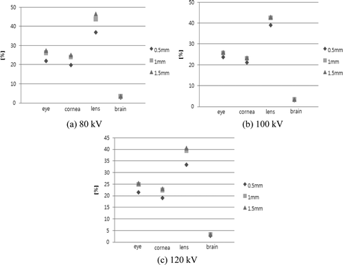
Figure 2. Absorbed dose reduction rate before and after use of an eye shield. (a) 80 kV. (b) 100 kV. (c) 120 kV.