Figures & data
Figure 1 Examples of (a) intensity distribution of neutrons and gamma rays and (b) neutron energy spectrum within the RPV in a PWR (figures drawn using radiation transport calculation data from [Citation8])
![Figure 1 Examples of (a) intensity distribution of neutrons and gamma rays and (b) neutron energy spectrum within the RPV in a PWR (figures drawn using radiation transport calculation data from [Citation8])](/cms/asset/3f25b3d3-c6cc-48a9-ae48-9bd865644725/tnst_a_772448_o_f0001g.gif)
Figure 2 Maximum fluence or dose and typical temperature for 40-year operation of a reactor pressure vessel and core internals
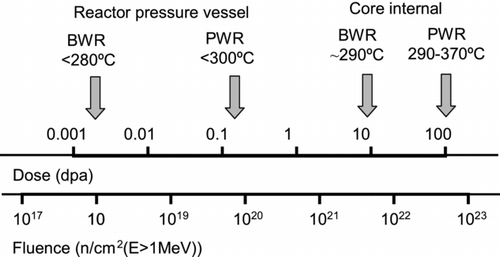
Figure 3 Schematic embrittlement trends in (a) low-flux BWR and (b) high-flux PWR conditions
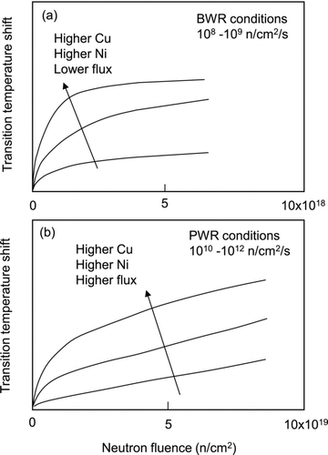
Figure 4 Comparison of transition temperature shifts between surveillance data and MTR data: (a) high-Cu BWR surveillance material and (b) low-Cu PWR surveillance material. The data from references [Citation32,Citation33] are replotted and trends are shown
![Figure 4 Comparison of transition temperature shifts between surveillance data and MTR data: (a) high-Cu BWR surveillance material and (b) low-Cu PWR surveillance material. The data from references [Citation32,Citation33] are replotted and trends are shown](/cms/asset/7d69ac8b-db90-4227-a0bf-7c4f2603fd63/tnst_a_772448_o_f0004g.gif)
Figure 5 Examples of 3DAP atom maps showing (a) Cu-rich clusters and (b) Mn–Ni–Si clusters in A533B steels irradiated with heavy ions to 1 dpa at 290ºC (courtesy of Katsuhiko Fujii of the Institute of Nuclear Safety System, Inc.). It should be noted that Mn–Ni–Si clusters are also found in (a)
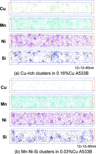
Figure 6 CDB spectra in 0.12%Cu A533B steels irradiated in a MTR at 290°C [Citation37]
![Figure 6 CDB spectra in 0.12%Cu A533B steels irradiated in a MTR at 290°C [Citation37]](/cms/asset/4ce08256-78f5-4f06-ae40-8f585a5d6d30/tnst_a_772448_o_f0006g.gif)
Figure 7 3DAP data of density and diameter for solute clusters in MTR-irradiated A533B steels with various Cu and Ni contents [Citation77]. Reprinted, with permission, from the Journal of ASTM International, Volume 6, Issue 7, copyright ASTM International, 100 Barr Harbor Drive, West Conshohocken, PA 19428
![Figure 7 3DAP data of density and diameter for solute clusters in MTR-irradiated A533B steels with various Cu and Ni contents [Citation77]. Reprinted, with permission, from the Journal of ASTM International, Volume 6, Issue 7, copyright ASTM International, 100 Barr Harbor Drive, West Conshohocken, PA 19428](/cms/asset/f7f7f542-959c-4280-8f7f-110147427d6d/tnst_a_772448_o_f0007g.jpg)
Figure 8 Relationship between cluster volume fraction (V f 1/2) and transition temperature shift (ΔRT NDT) in surveillance specimens [Citation59]. Reprinted, with permission, from the Journal of ASTM International, Volume 7, Issue 3, copyright ASTM International, 100 Barr Harbor Drive, West Conshohocken, PA 19428
![Figure 8 Relationship between cluster volume fraction (V f 1/2) and transition temperature shift (ΔRT NDT) in surveillance specimens [Citation59]. Reprinted, with permission, from the Journal of ASTM International, Volume 7, Issue 3, copyright ASTM International, 100 Barr Harbor Drive, West Conshohocken, PA 19428](/cms/asset/7229f107-c90c-4607-a964-31780b437672/tnst_a_772448_o_f0008g.gif)
Figure 9 Effect of dose rate on the number of vacancy jumps from kinetic Monte Carlo calculations [Citation94]
![Figure 9 Effect of dose rate on the number of vacancy jumps from kinetic Monte Carlo calculations [Citation94]](/cms/asset/9e25072c-1cd7-426a-bf87-46d308274cbd/tnst_a_772448_o_f0009g.gif)
Figure 10 TEM images showing interstitial dislocation loops in A533B steels irradiated with heavy ions to 1 dpa at 290°C: weak-beam images with diffraction vectors (a) g = 011 and (b) g = 200 close to the [011] pole [Citation98]
![Figure 10 TEM images showing interstitial dislocation loops in A533B steels irradiated with heavy ions to 1 dpa at 290°C: weak-beam images with diffraction vectors (a) g = 011 and (b) g = 200 close to the [011] pole [Citation98]](/cms/asset/98571c13-54d4-4e01-bec0-e834e864aa51/tnst_a_772448_o_f0010g.gif)
Figure 11 Positron lifetime data in weld surveillance specimens (0.13%Cu for Doel-1 and 0.30%Cu for Doel-2) [Citation53]
![Figure 11 Positron lifetime data in weld surveillance specimens (0.13%Cu for Doel-1 and 0.30%Cu for Doel-2) [Citation53]](/cms/asset/fc2b7042-27cc-4a7c-9efe-3a9c9f1cecb2/tnst_a_772448_o_f0011g.gif)
Figure 12 Irradiation hardening (Δσy ) versus transition temperature shift (ΔT 41J) in MTR-irradiated A533B steels with various levels of P segregation at GBs (ΔCP gb) [Citation121]. Reprinted, with permission, from the Journal of ASTM International, Volume 6, Issue 7, copyright ASTM International, 100 Barr Harbor Drive, West Conshohocken, PA 19428
![Figure 12 Irradiation hardening (Δσy ) versus transition temperature shift (ΔT 41J) in MTR-irradiated A533B steels with various levels of P segregation at GBs (ΔCP gb) [Citation121]. Reprinted, with permission, from the Journal of ASTM International, Volume 6, Issue 7, copyright ASTM International, 100 Barr Harbor Drive, West Conshohocken, PA 19428](/cms/asset/9bc71266-7210-4e7f-a33c-a64512d55710/tnst_a_772448_o_f0012g.gif)
Figure 13 Plots of master curve reference temperature shift ΔT 0 versus Charpy transition temperature shift ΔT 41J for A533B steel data sets containing high-shift data. The dotted line shows the correlation given by Sokolov and Nanstad [Citation148]
![Figure 13 Plots of master curve reference temperature shift ΔT 0 versus Charpy transition temperature shift ΔT 41J for A533B steel data sets containing high-shift data. The dotted line shows the correlation given by Sokolov and Nanstad [Citation148]](/cms/asset/90578703-3216-43bf-ae5f-c05e57893ba4/tnst_a_772448_o_f0013g.gif)
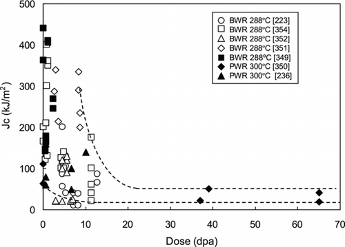
Figure 14 Constant load SCC test data in the stress ratio to yield strength (σ/σy ) versus dose maps in (a) BWR water conditions (288°C, 32 ppm DO) and PWR water conditions (320–340°C, 2.7 ppm DH)
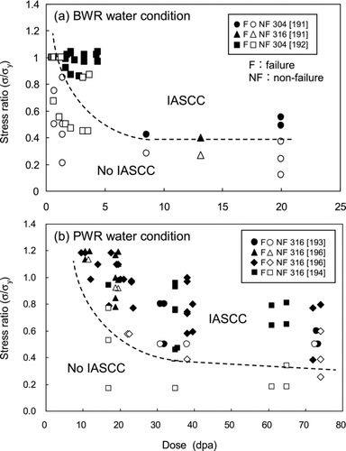
Figure 15 Dose dependence of CGR in BWR water conditions at both high ECP (>150 mVSHE) and low ECP (<–200 mVSHE)
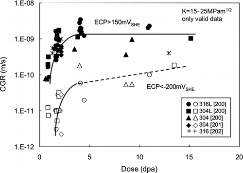
Figure 16 CGR data of 304 SSs from in-pile tests and PIEs in BWR NWC water conditions [Citation208]
![Figure 16 CGR data of 304 SSs from in-pile tests and PIEs in BWR NWC water conditions [Citation208]](/cms/asset/ce51e8fc-3dcf-4483-b88f-47580eda9db7/tnst_a_772448_o_f0016g.jpg)
Figure 17 TEM images showing microstructural features observed in CW type 316 SSs PWR-irradiated to 53 dpa: (a) rel-rod dark field image of dislocation loops; (b) dark field image of dislocation loops and black dots; (c) dark field image of Ni3Si precipitates; and (d) defocused image of bubbles [Citation220]
![Figure 17 TEM images showing microstructural features observed in CW type 316 SSs PWR-irradiated to 53 dpa: (a) rel-rod dark field image of dislocation loops; (b) dark field image of dislocation loops and black dots; (c) dark field image of Ni3Si precipitates; and (d) defocused image of bubbles [Citation220]](/cms/asset/8c973cd3-ec29-4523-9bda-f13a3e248c87/tnst_a_772448_o_f0017g.gif)
Figure 18 Change in (a) yield strength and (b) fracture elongation in LWR-irradiated SSs
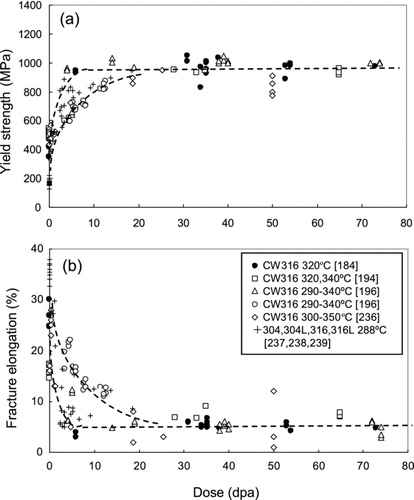
Figure 19 Near-surface deformation microstructure in CW type 316 SSs deformed to 3% at 320°C: (a) irradiated to 35 dpa showing coarse slips and surface steps with an enlarged image of the dislocation channel and (b) unirradiated showing tangled dislocations [Citation245]
![Figure 19 Near-surface deformation microstructure in CW type 316 SSs deformed to 3% at 320°C: (a) irradiated to 35 dpa showing coarse slips and surface steps with an enlarged image of the dislocation channel and (b) unirradiated showing tangled dislocations [Citation245]](/cms/asset/a49ab343-49c7-4276-bafb-1e763d9411fb/tnst_a_772448_o_f0019g.gif)
Figure 20 Schematic illustration depicting (a) three types of interactions of dislocation channels with GBs; (b) SEM image showing dislocation pileups in irradiated CW type 316 SSs after slow deformation to 13% at 320°C; and (c) TEM image showing channel transfer in irradiated CW type 316 SSs after slow deformation to ∼3% at 320°C
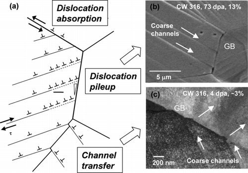
Figure 21 Typical solute distributions across a GB and changes in GB segregation of Cr, Ni and Si with dose in LWR-irradiated SSs
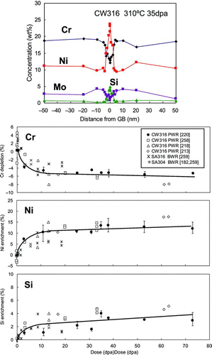
Figure 22 Data of stress relaxation in type 304 and 316L SSs under MTR irradiation at 288°C
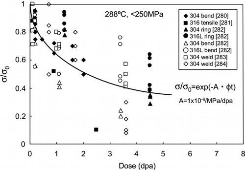
Figure 23 Dose-dependent changes in IASCC, mechanical properties and RIS in PWR-irradiated CW type 316 SSs [Citation298]
![Figure 23 Dose-dependent changes in IASCC, mechanical properties and RIS in PWR-irradiated CW type 316 SSs [Citation298]](/cms/asset/4d0ea4a0-e959-484a-aa45-09be967fb43d/tnst_a_772448_o_f0023g.gif)
Figure 24 TEM image and solute distribution near the IASCC crack tip in CW type 316 SSs PWR-irradiated to 38 dpa after the constant load test at 750 MPa in PWR water conditions [Citation304]
![Figure 24 TEM image and solute distribution near the IASCC crack tip in CW type 316 SSs PWR-irradiated to 38 dpa after the constant load test at 750 MPa in PWR water conditions [Citation304]](/cms/asset/a43d6a87-a4f6-4332-ab81-56ab65713ae8/tnst_a_772448_o_f0024g.jpg)
Figure 25 Schematic illustration depicting various processes related to IASCC
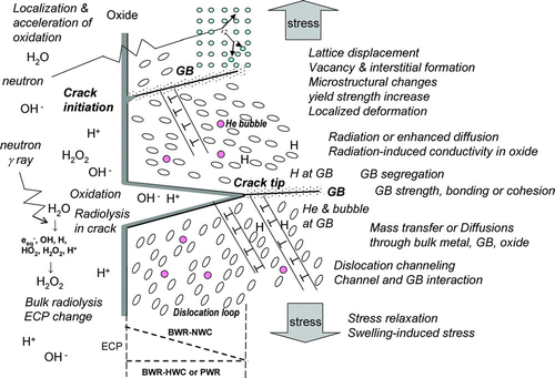
Figure 26 IG cracks formed in PWR-irradiated CW type 316 SSs after slow deformation to ∼3% at 300°C in an argon atmosphere [Citation245]
![Figure 26 IG cracks formed in PWR-irradiated CW type 316 SSs after slow deformation to ∼3% at 300°C in an argon atmosphere [Citation245]](/cms/asset/ffa7cfd3-2a5e-4754-ab2d-8d910a4246b7/tnst_a_772448_o_f0026g.gif)
Figure 27 Contribution of localized deformation as measured by the weighted average channel height to cracking in SSs irradiated with protons to 1–5 dpa and SCC-tested in BWR water conditions [Citation247]
![Figure 27 Contribution of localized deformation as measured by the weighted average channel height to cracking in SSs irradiated with protons to 1–5 dpa and SCC-tested in BWR water conditions [Citation247]](/cms/asset/0ee57a10-5726-401a-b6f2-4ca0ccb19427/tnst_a_772448_o_f0027g.jpg)
Figure 28 Distribution of Fe, Ni, Cr and O in the cross section of the surface oxide layer in type 316 SSs PWR-irradiated to 20 dpa after immersion in PWR water conditions for 1000 h [Citation322]
![Figure 28 Distribution of Fe, Ni, Cr and O in the cross section of the surface oxide layer in type 316 SSs PWR-irradiated to 20 dpa after immersion in PWR water conditions for 1000 h [Citation322]](/cms/asset/9fca3416-f1f3-4e0e-855c-e4efaccb74c6/tnst_a_772448_o_f0028g.jpg)
Figure 29 Swelling data in various SSs irradiated at LWR-relevant temperatures of 290–390ºC
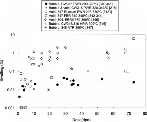
Figure 30 Fracture toughness data in LWR-irradiated SSs tested at ∼300ºC