Figures & data
Table 1. Isolates examined in this study.
Figure 1. Phylogeny of members of the Leotiomycetes inferred from ML analysis of the concatenated nuc 18S + nuc 28S + mt 18S + RPB2 (four-gene) data set. An asterisk (*) indicates branches with ML BS = 100% and PP values = 1.0. Branch support in nodes ≥70% ML BS and ≥0.90 PP is indicated above or below branches. “T” after an accession number indicates that the strain is an ex-type culture; “#” after an accession number indicates that the strain has demonstrated antifungal activity. Names in bold are taxonomic novelties. Shading is used to differentiate lineages and highlight taxa of interest.
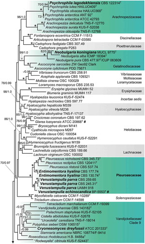
Figure 1. (Continued).
Figure 2. Phylogeny of the Pleuroascaceae inferred from ML analysis of the concatenated ITS + nuc 18S + nuc 28S + mt 18S + RPB2 (five-gene) data set. Details as in .
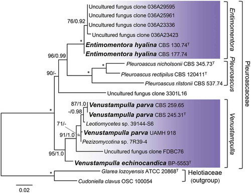
Figure 3. Position of Neobulgaria koningiana (MUCL 9775) within Neobulgaria based on ML analysis of ITS sequences. Details as in .
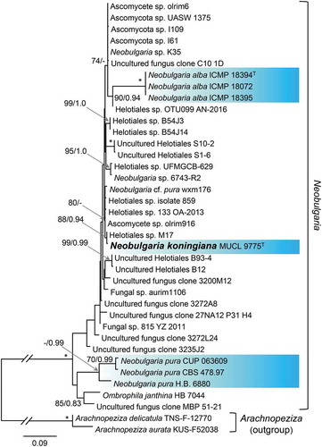
Figure 4. Phylogeny of Psychrophila inferred from ML analysis of ITS sequences. Details as in .
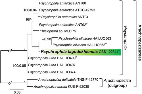
Figure 5. Position of Cryonesomyces dreyfussii (ATCC 201333) within the Vandijckellaceae/Clade 9 inferred from ML analysis of the concatenated ITS + nuc 18S + nuc 28S + RPB2 data set. Details as in .
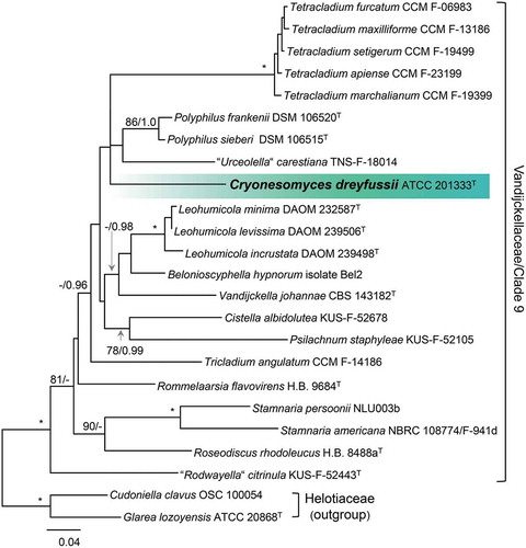
Figure 6. Colonies of species of Entimomentora and Pleuroascus on CBSOA, MLA, OA, and PCA after 28 d. A. E. hyalina (ex-type CBS 130.74). B. Pl. nicholsonii (ex-type CBS 345.73). C. Pl. rectipilus (ex-type CBS 120411). D. Pl. rilstonii (CBS 537.74).
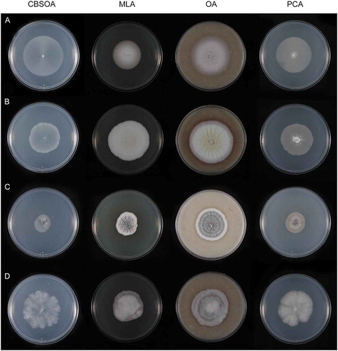
Figure 7. Entimomentora hyalina. A–G. Phialides with conidia (MLA slide culture, 13–17 d). H. Conidia (MLA, 10 d). A–D, H from CBS 177.74; E–G from CBS 130.74. Bars = 10 μm.
Figure 8. Colonies of species of Venustampulla on CBSOA, MLA, OA, and PCA after 28 d. A. V. echinocandica (ex-type BP-5553). B. V. parva (CBS 245.31). C. V. parva (CBS 259.65). D. V. parva (ex-type of Scopulariopsis parvula UAMH 918).
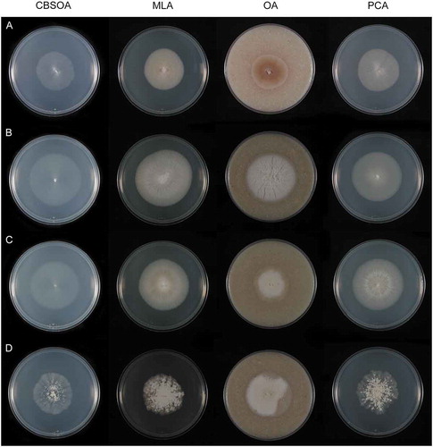
Figure 9. Venustampulla echinocandica (ex-type BP-5553). A–E. Phialides with conidia in chains; arrow indicates a percurrently proliferating phialide (MLA slide cultures, 11 d). F. Conidia (PCA, 12 d). Bars = 10 μm.
Figure 10. Venustampulla parva. A–F. Phialides with conidia (MLA slide cultures, 10–11 d). G. Conidia (MLA, 15 d). A–D, F from CBS 259.65; E, G from ex-type of Paecilomyces parvus CBS 245.31. Bars = 10 μm.
Figure 11. Venustampulla parva (ex-type of Scopulariopsis parvula UAMH 918). A–F. Phialides with conidia (MLA slide cultures, 10–15 d). G. Conidia (MLA, 15 d). Bars = 10 μm.
Figure 12. Colonies of Neobulgaria and Cryonesomyces on CBSOA, MLA, OA, and PCA. A. Neobulgaria koningiana (ex-type MUCL 9775) after 14 d. B. Cryonesomyces dreyfussii (ex-type ATCC 201333) after 28 d.
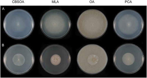
Figure 13. Cryonesomyces dreyfussii (ex-type ATCC 20133). A–F. Phialides with conidia (MLA slide culture, 13 d). G, H. Conidia (MLA slide culture, 13 d). Bars = 10 μm.
Figure 14. Neobulgaria koningiana (ex-type MUCL 9775). A–G. Phialides with conidia (MLA slide cultures, 10 d). H–J. Conidia (MLA, 10 d). Bars = 10 μm.
Figure 15. Colonies of Psychrophila on CBSOA, MLA, OA, and PCA after 28 d. A. Ps. lagodekhiensis (ex-type CBS 122314). B. Ps. antarctica (ATCC 42793).
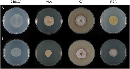
Figure 16. Colonies of Psychrophila on PDA after 28 d at 4, 10, and 20 C. A. Ps. lagodekhiensis (ex-type CBS 122314). B. Ps. antarctica (ATCC 42793).
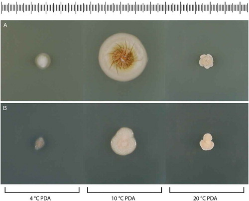
Figure 17. Psychrophila antarctica (ATCC 42793). A–K. Phialides with conidia (MLA slide culture, 13 d). Bars = 10 μm.
Figure 18. Psychrophila lagodekhiensis (ex-type CBS 122314). A–K. Phialides with conidia (MLA slide culture, 13 d). L. Conidia (MLA slide culture, 13 d). Bars = 10 μm.