Figures & data
Figure 1 Cololejeunea grossepapillosa. A, Ventral view of leaf. B, Dorsal view of leaf. C, Athecal vegetative branch. D, Stem sections. E, Ventral view of shoot. F, Ventral view of leaf showing gemma initial cells. G, Gemmae. H, Gemmaling. I, Dorsal view of leaf showing gemma initial cells, papillae omitted. Scale bar A–D, F–I: 100 µm; E: 250 µm.
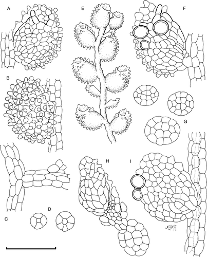
Table 1 Characters comparing Cololejeunea grossepapillosa with known New Zealand species with which it could be confused.
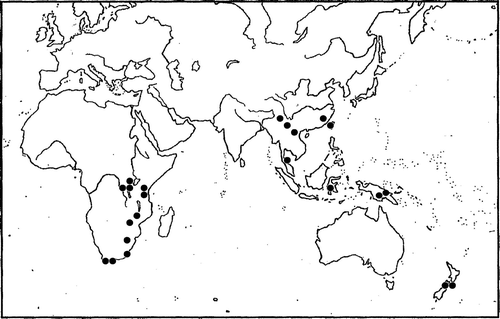
Figure 2 Worldwide distribution of Cololejeunea grossepapillosa, showing disjunction between eastern and southern Africa, Asia and New Zealand. Black dots represent the approximate location of known sightings and herbarium records. The two South Island records are reported here.