707
Views
5
CrossRef citations to date
0
Altmetric
Articles

Cavernosa kapitiana (Bacillariophyceae): biogeography and morphology of the different life cycle stages

, &
Pages 443-459 | Received 15 Dec 2010, Accepted 11 Apr 2011, Published online: 21 Nov 2011

Figures & data

Table 1  Morphological characteristics of Cavernosa and similar genera.

Figure 1 Map of the investigated locations. A, Part of the southern hemisphere with the locations of the Crozet Archipelago and New Zealand. B, Ile de la Possession (Crozet Archipelago) with the locations where Cavernosa was found. C, New Zealand showing the position of Kapiti Island.
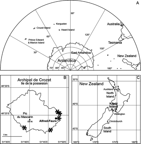
Figure 1  Map of the investigated locations. A, Part of the southern hemisphere with the locations of the Crozet Archipelago and New Zealand. B, Ile de la Possession (Crozet Archipelago) with the locations where Cavernosa was found. C, New Zealand showing the position of Kapiti Island.

Table 2  Material of Cavernosa kapitiana examined for this contribution.

Figure 2 Chain forming (A–C) and plastids (A,D,E) in Cavernosa kapitiana from Ile de la Possession. A–C, Chains linked by spines showing multiple plastids per cell. The arrows in C indicate the position of linking spines. D,E, Position of the caverns indicated by arrows. Note also the large number of discoid plastids. Scale bars, 20 μm (A,B), 10 μm (C–E).
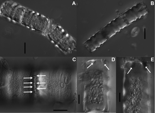
Figure 2  Chain forming (A–C) and plastids (A,D,E) in Cavernosa kapitiana from Ile de la Possession. A–C, Chains linked by spines showing multiple plastids per cell. The arrows in C indicate the position of linking spines. D,E, Position of the caverns indicated by arrows. Note also the large number of discoid plastids. Scale bars, 20 μm (A,B), 10 μm (C–E).

Figure 3  Light micrographs of Cavernosa kapitiana from Ile de la Possession. A,B, Entire frustules showing the zonation on the mantle and the presence of spines and caverns (indicated by arrows). C,D, Two micrographs of the same valve at different focus level showing the surface structure (C), the caverns (D) and the rimoportula in the valve centre (D, arrow). Scale bars, 10 μm.
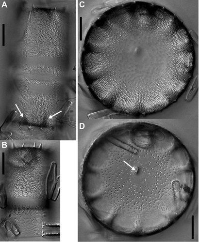
Figure 3  Light micrographs of Cavernosa kapitiana from Ile de la Possession. A,B, Entire frustules showing the zonation on the mantle and the presence of spines and caverns (indicated by arrows). C,D, Two micrographs of the same valve at different focus level showing the surface structure (C), the caverns (D) and the rimoportula in the valve centre (D, arrow). Scale bars, 10 μm.

Figure 4  Scanning electron micrographs of Cavernosa kapitiana from Ile de la Possession. A, Concave valve face. B, Convex valve face. C, Detail of the surface structure of a concave valve showing three different ornamentation elements: pores, granules and ridges. The arrow indicates the position of the rimoportula. D, Surface structure of the valve mantle. E, Detailed valve face view of the stellate spines. F, Valve–mantle transition with the position of the spines. Scale bars, 10 μm except for (C,E), 1 μm.
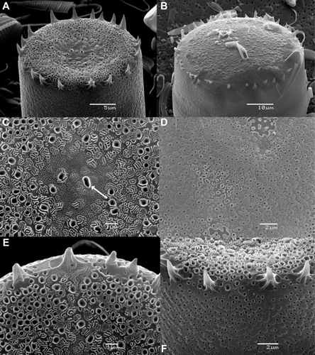
Figure 4  Scanning electron micrographs of Cavernosa kapitiana from Ile de la Possession. A, Concave valve face. B, Convex valve face. C, Detail of the surface structure of a concave valve showing three different ornamentation elements: pores, granules and ridges. The arrow indicates the position of the rimoportula. D, Surface structure of the valve mantle. E, Detailed valve face view of the stellate spines. F, Valve–mantle transition with the position of the spines. Scale bars, 10 μm except for (C,E), 1 μm.

Figure 5 Scanning electron micrographs of the girdle structure of Cavernosa kapitiana from Ile de la Possession. A, Entire mantle and girdle structure with the typical zonation. The different parts of the mantle and the girdle are indicated. The area in the black circle shows the structure of the valvocopula under the mantle. B, Detail of the valvocopula with the fimbriate pars interior and the pars exterior. C, Slit of a copula in which the ligula of the following abvalvar copula fits. D, Interior view of the girdle. Scale bars, 10 μm.
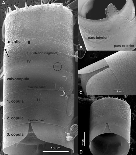
Figure 5  Scanning electron micrographs of the girdle structure of Cavernosa kapitiana from Ile de la Possession. A, Entire mantle and girdle structure with the typical zonation. The different parts of the mantle and the girdle are indicated. The area in the black circle shows the structure of the valvocopula under the mantle. B, Detail of the valvocopula with the fimbriate pars interior and the pars exterior. C, Slit of a copula in which the ligula of the following abvalvar copula fits. D, Interior view of the girdle. Scale bars, 10 μm.

Figure 6 Scanning electron micrographs of the internal valve and rimoportulae of Cavernosa kapitiana from Ile de la Possession. A, Valve interior showing the caverns and one central rimoportula. B, Presence of two rimoportulae on a valve emerging from the first division of an initial cell. The arrows indicate the ringleiste. C,D, Structure of the rimportula seen from different angles. E, Detail of the two rimoportulae in (B). Scale bars, 10 μm except for (C, D), 1 μm.
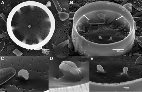
Figure 6  Scanning electron micrographs of the internal valve and rimoportulae of Cavernosa kapitiana from Ile de la Possession. A, Valve interior showing the caverns and one central rimoportula. B, Presence of two rimoportulae on a valve emerging from the first division of an initial cell. The arrows indicate the ringleiste. C,D, Structure of the rimportula seen from different angles. E, Detail of the two rimoportulae in (B). Scale bars, 10 μm except for (C, D), 1 μm.

Figure 7 Light and scanning electron micrographs of a globular initial cell of Cavernosa kapitiana from Ile de la Possession. A, LM view of complete initial cell. B, SEM view of a complete initial cell. C,D, SEM view of external surface structure of an initial valve. A small ringleiste is visible on (D). E, SEM internal view of an initial valve with the presence of two rimoportulae and only weakly developed caverns. F, SEM detail of the internal surface structure showing an irregular pattern of areolae openings and two rimportulae. Scale bars, 10 μm except for (F), 5 μm.
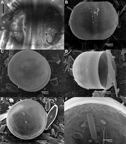
Figure 7  Light and scanning electron micrographs of a globular initial cell of Cavernosa kapitiana from Ile de la Possession. A, LM view of complete initial cell. B, SEM view of a complete initial cell. C,D, SEM view of external surface structure of an initial valve. A small ringleiste is visible on (D). E, SEM internal view of an initial valve with the presence of two rimoportulae and only weakly developed caverns. F, SEM detail of the internal surface structure showing an irregular pattern of areolae openings and two rimportulae. Scale bars, 10 μm except for (F), 5 μm.

Figure 8 Light micrographs of the cell following the first division of an initial cell of Cavernosa kapitiana from Ile de la Possession. A, Girdle view of a cell with a globular valve and a flat valve. B, Face view of a flat valve with two rimoportulae in the valve centre (arrows). Scale bar, 10 μm.
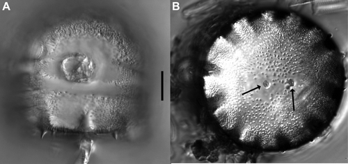
Figure 8  Light micrographs of the cell following the first division of an initial cell of Cavernosa kapitiana from Ile de la Possession. A, Girdle view of a cell with a globular valve and a flat valve. B, Face view of a flat valve with two rimoportulae in the valve centre (arrows). Scale bar, 10 μm.

Figure 9 Light and Scanning electron micrographs of a bell-shaped initial cell of Cavernosa kapitiana from Ile de la Possession. A, LM view of a bell-shaped initial cell. B, SEM view of a bell-shaped initial valve. C, SEM view of external detail of the handle part of the bell-shaped initial valve. Spines are clearly absent and the surface ornamentation is irregular. D, SEM internal view of a bell-shaped initial valve. Scale bars, 10 μm except for (C), 2 µm.
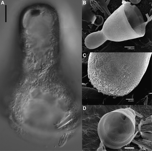
Figure 9  Light and Scanning electron micrographs of a bell-shaped initial cell of Cavernosa kapitiana from Ile de la Possession. A, LM view of a bell-shaped initial cell. B, SEM view of a bell-shaped initial valve. C, SEM view of external detail of the handle part of the bell-shaped initial valve. Spines are clearly absent and the surface ornamentation is irregular. D, SEM internal view of a bell-shaped initial valve. Scale bars, 10 μm except for (C), 2 µm.

Figure 10 Light (A,B) and scanning electron (C–E) micrographs of Cavernosa kapitiana from the type population on Kapiti Island, New Zealand. A, LM valve view with the presence of a rimoportula (arrow) and the typical caverns. B, LM girdle view showing the mantle and part of the girdle. C, SEM picture of the mantle. Note the comparably small spines. D, SEM valve face view of a concave valve with the opening of the rimoportula (arrow) and the typically ornamented surface structure. E, SEM internal view with the position of the rimoportula (arrow) and the caverns. Scale bars, 10 μm.
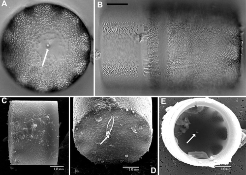
Figure 10  Light (A,B) and scanning electron (C–E) micrographs of Cavernosa kapitiana from the type population on Kapiti Island, New Zealand. A, LM valve view with the presence of a rimoportula (arrow) and the typical caverns. B, LM girdle view showing the mantle and part of the girdle. C, SEM picture of the mantle. Note the comparably small spines. D, SEM valve face view of a concave valve with the opening of the rimoportula (arrow) and the typically ornamented surface structure. E, SEM internal view with the position of the rimoportula (arrow) and the caverns. Scale bars, 10 μm.

Table 3  Morphometric and morphological comparison of Cavernosa kapitiana from the type locality (Kapiti Island, New Zealand) and Ile de la Possession (southern Indian Ocean). n=x refers to number of measured specimens.

Reprints and Corporate Permissions

Please note: Selecting permissions does not provide access to the full text of the article, please see our help page How do I view content?

To request a reprint or corporate permissions for this article, please click on the relevant link below:

Academic Permissions

Please note: Selecting permissions does not provide access to the full text of the article, please see our help page How do I view content?

Obtain permissions instantly via Rightslink by clicking on the button below:

If you are unable to obtain permissions via Rightslink, please complete and submit this Permissions form. For more information, please visit our Permissions help page.