7,061
Views
32
CrossRef citations to date
0
Altmetric
Research articles

Non-glandular trichomes in Lamiaceae and Verbenaceae species: morphological and histochemical features indicate more than physical protection

, &
Pages 446-457 | Received 22 Apr 2016, Accepted 14 Jun 2016, Published online: 25 Jul 2016

Figures & data

Figure 1. Scanning electron micrographs of the leaf blade in Lamiaceae and Verbenaceae species. A, B, Abaxial and adaxial leaf surfaces, respectively, in Lippia origanoides showing non-glandular trichomes. C–J, Morphological features of the non-glandular trichomes in Verbenaceae and Lamiaceae species. C, Aegiphila verticillata. Detail showing the basal cells. D, Hyptis villosa. E, F, Plectranthus barbatus. G, Lantana camara. Observe accumulations of secretion (detail) on the apical cell. H, Lippia alba. I, Lippia origanoides. J, Stachytarpheta cayennensis. Scale bars: A, B = 500 µm; C, D, F, J = 50 µm; E = 200 µm; G–I = 25 µm.

Figure 1. Scanning electron micrographs of the leaf blade in Lamiaceae and Verbenaceae species. A, B, Abaxial and adaxial leaf surfaces, respectively, in Lippia origanoides showing non-glandular trichomes. C–J, Morphological features of the non-glandular trichomes in Verbenaceae and Lamiaceae species. C, Aegiphila verticillata. Detail showing the basal cells. D, Hyptis villosa. E, F, Plectranthus barbatus. G, Lantana camara. Observe accumulations of secretion (detail) on the apical cell. H, Lippia alba. I, Lippia origanoides. J, Stachytarpheta cayennensis. Scale bars: A, B = 500 µm; C, D, F, J = 50 µm; E = 200 µm; G–I = 25 µm.

Table 1. Density of glandular and non-glandular trichomes (mm2) in leaves of Lamiaceae and Verbenaceae species.

Figure 2. Histochemical tests of non-glandular trichomes in leaves of Lamiaceae and Verbenaceae species. A–C, Aegiphila verticillata. D, E, Hyptis villosa. F–I, Plectranthus barbatus. J, K, Lantana camara. L, Lippia alba. M, N, Lippia origanoides. O–R, Stachytarpheta cayennensis. A, N, Q, Dragendorff reagent. B, G, O, Bromophenol blue. C, F, J, L, P, Ferric chloride. D, H, K, Sudan IV. Arrows indicate oil droplets on the cell walls. E, I, R, Nadi reagent. M, Periodic acid/Schiff reagent. Scale bars = 50 µm.

Figure 2. Histochemical tests of non-glandular trichomes in leaves of Lamiaceae and Verbenaceae species. A–C, Aegiphila verticillata. D, E, Hyptis villosa. F–I, Plectranthus barbatus. J, K, Lantana camara. L, Lippia alba. M, N, Lippia origanoides. O–R, Stachytarpheta cayennensis. A, N, Q, Dragendorff reagent. B, G, O, Bromophenol blue. C, F, J, L, P, Ferric chloride. D, H, K, Sudan IV. Arrows indicate oil droplets on the cell walls. E, I, R, Nadi reagent. M, Periodic acid/Schiff reagent. Scale bars = 50 µm.

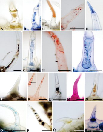
Table 2. Histochemistry of the non-glandular trichomes in leaves of Lamiaceae and Verbenaceae species.

Figure 3. Transmission electron micrographs of the non-glandular trichomes in leaves of two Verbenaceae and one Lamiaceae species. A–E, Lantana camara. A, Apical cell showing nucleus with irregular contour and plastids (Pl) with starch grains and voluminous oil bodies (Ol) in the cytoplasm. Va: vacuole. B, Detail showing lipidic substances (arrows) impregnated in the wall of the apical cell. C, Portion of apical cell showing mitochondria (Mi), smooth endoplasmic reticulum (Sr) and vesicles (Ve). Va: vacuole. D, Detail of apical cell with large plastids containing voluminous oil drops (Ol) and starch grains (St). Gb: Golgi body; Mi: mitochondria; Va: vacuole. E, Plasmodesmata (arrowhead) connecting apical (AC) and basal (BC) cells. Ol: oil drop; Sr: smooth endoplasmic reticulum; Va: vacuole; Ve: vesicles. F, G, Lippia origanoides. F, Apical cell. Observe thick cuticle (Ct) with electron-dense ramifications. Cw: cell wall; Gb: Golgi body; Mi: mitochondria; Ol: oil; Va: vacuole. G, Portion of basal cell exhibiting thick cell wall (Cw) with lamellar aspect and accumulations of electron-dense material (*) in the outer portion. Observe flocculate cytoplasm and oil (Ol) drops. H–J, Hyptis villosa. H, Apical cell with Golgi bodies (Gb) hyperactive in the vesicle production in the cytoplasm and paramural bodies (Pb) in the wide periplasmic space. Cw: cell wall. Nu: nucleus. Va: vacuole. I, Detail of apical (AC) and basal (BC) cells. Cw: cell wall. Gb: Golgi body. Mi: mitochondria. Va: vacuole. Observe vesicles (Ve) next to the plasmalemma in the basal cells. J, Portions of apical (AC) and basal (BC) cells with polyribosomes, rough endoplasmic reticulum (Rr) and vesicles (Ve). Va: vacuole. Scale bars = 1 μm.

Figure 3. Transmission electron micrographs of the non-glandular trichomes in leaves of two Verbenaceae and one Lamiaceae species. A–E, Lantana camara. A, Apical cell showing nucleus with irregular contour and plastids (Pl) with starch grains and voluminous oil bodies (Ol) in the cytoplasm. Va: vacuole. B, Detail showing lipidic substances (arrows) impregnated in the wall of the apical cell. C, Portion of apical cell showing mitochondria (Mi), smooth endoplasmic reticulum (Sr) and vesicles (Ve). Va: vacuole. D, Detail of apical cell with large plastids containing voluminous oil drops (Ol) and starch grains (St). Gb: Golgi body; Mi: mitochondria; Va: vacuole. E, Plasmodesmata (arrowhead) connecting apical (AC) and basal (BC) cells. Ol: oil drop; Sr: smooth endoplasmic reticulum; Va: vacuole; Ve: vesicles. F, G, Lippia origanoides. F, Apical cell. Observe thick cuticle (Ct) with electron-dense ramifications. Cw: cell wall; Gb: Golgi body; Mi: mitochondria; Ol: oil; Va: vacuole. G, Portion of basal cell exhibiting thick cell wall (Cw) with lamellar aspect and accumulations of electron-dense material (*) in the outer portion. Observe flocculate cytoplasm and oil (Ol) drops. H–J, Hyptis villosa. H, Apical cell with Golgi bodies (Gb) hyperactive in the vesicle production in the cytoplasm and paramural bodies (Pb) in the wide periplasmic space. Cw: cell wall. Nu: nucleus. Va: vacuole. I, Detail of apical (AC) and basal (BC) cells. Cw: cell wall. Gb: Golgi body. Mi: mitochondria. Va: vacuole. Observe vesicles (Ve) next to the plasmalemma in the basal cells. J, Portions of apical (AC) and basal (BC) cells with polyribosomes, rough endoplasmic reticulum (Rr) and vesicles (Ve). Va: vacuole. Scale bars = 1 μm.

Reprints and Corporate Permissions

Please note: Selecting permissions does not provide access to the full text of the article, please see our help page How do I view content?

To request a reprint or corporate permissions for this article, please click on the relevant link below:

Academic Permissions

Please note: Selecting permissions does not provide access to the full text of the article, please see our help page How do I view content?

Obtain permissions instantly via Rightslink by clicking on the button below:

If you are unable to obtain permissions via Rightslink, please complete and submit this Permissions form. For more information, please visit our Permissions help page.