Figures & data
Figure 1. Map of Cenozoic New Zealand localities with recorded leaf fragments (red) of cycad fossils with anastomosing venation (Pole Citation1992, Citation2007; this study) and Late Cretaceous to Cenozoic cycad pollen reports from FRED, Raine et al (Citation2011), or as unpublished reports (D.C. Mildenhall, 2017, pers. comm.): Cretaceous to Paleocene (green), Eocene (blue), Oligocene (yellow) and Miocene (pale purple). See Appendix 1 for pollen locality details.
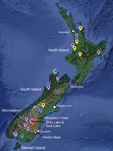
Figure 2. Pterostoma neehoffii sp. nov., all cuticle images derived from OU35425a. A, Holotype (OU35425a), showing anastomosing veins (arrows); B, Paratype counterpart (OU35425b); C, SEM of adaxial epidermis showing smooth outer surface without obvious cellular detail or hairs; D, SEM of adaxial epidermis inner surface showing strongly sinuous anticlinal walls with buttressing; E, TLM of abaxial surface showing intercostal stomatal band, elongated costal cells and two crystalliferous cells along the vein (arrow); F, TLM of abaxial intercostal cells showing randomly oriented stomata and buttressing of sinuous anticlinal walls; G, SEM of abaxial intercostal cells showing sparse cuticular ridges, sunken stomata and prominent stomal rings; H, TLM of sunken stomate and stomatal ring (external view); I, SEM of stomate (internal view) showing guard cell flanges and thin-walled subsidiary cells. Scale bars A, B = 20 mm, C = 100 µm, D, F, G = 50 µm, E = 100 µm, H, I = 25 µm.
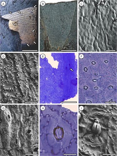
Figure 3. Comparative Eocene Australian Pterostoma fossils described by Hill (Citation1980). A–D, Pterostoma zamioides from Anglesea (P150055 and AN 2284), Victoria, E, F, P. anastomans from Nerriga, New South Wales (N0157); all samples held at the Melbourne Museum. A, Pinna fragment (AN 2284) showing anastomosing veins (arrows); B, SEM of P150055 adaxial surface showing strongly sinuous anticlinal cell walls; C, SEM of P150055 stomatal ring and cuticular ridges; D, SEM of P150055 stomate (interior view) showing guard cell flanges; E, Pinna fragment of distal pinna fragment of N0157 showing anastomosing veins (arrows); F, SEM of N0157 intercostal cells showing cuticular ridges, a trichome base (arrow) and sunken stomata with prominent rings; G, Cycad-like ?Avicennia diaspore (OU 30770) described from Landslip Hill by Campbell (Citation2002) showing short apical beak (arrow); H, Cycad-like ?Avicennia diaspore (OU 30771) described from Landslip Hill by Campbell (Citation2002) showing basal attachment scar (arrow); I, Cycas revoluta Thunb. seed showing short apical beak (white arrow) and basal attachment (hilum) scar (black arrow). Scale bars A, E = 2 mm B, F = 50 µm, C, D = 20 µm, G–I = 10 mm; photos A–F courtesy of Bob Hill.
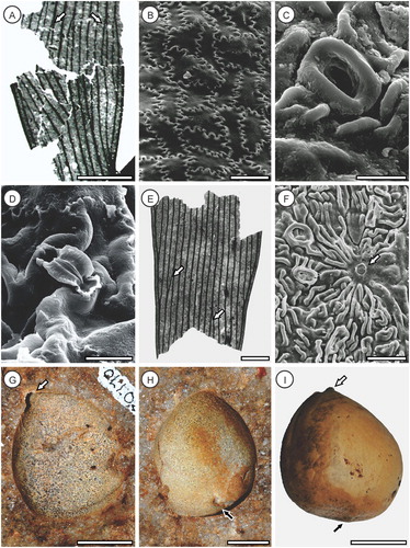
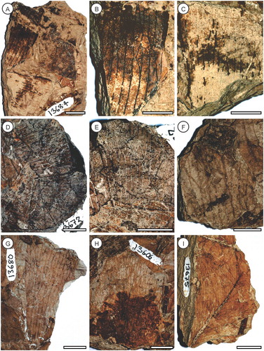
Figure 4. Photographs of specimens reported by Pole (Citation1992) as possible cycads from Bannockburn. A, OU13684, showing broad attachment of two pinnae to the rachis (arrow); B, C, details of same showing anastomosing veins; D, OU13672, possibly two pinna side by side; E, detail of same showing anastomosing veins; F, OU12583, fragment of pinna showing anastomosing veins; G, OU13680, proximal pinna fragment; H, OU13606, fragment of pinna showing anastomosing veins; I, OU13075, bases of two pinnae showing very broad attachment to the rachis (arrow). See Pole (Citation1992) for detailed drawings of the venation. Scale bars A–F, H, I = 10 mm, G = 5 mm.