Figures & data
Fig. 1 (A–G) Living Polydora haswelli: (A) whole body; (B) anterior lateral right body and palps; (C) posterior and pygidium; (D) egg capsule string; (E) anterior dorsal body; (F) palps and dorsal head; (G) head in right lateral view. (H–I) Living Polydora websteri: (H) anterior body and palps, with individual egg capsules; (I) posterior and pygidium in lateral view. (J) Preserved Polydora websteri palps groove-edge pigmentation. Scale bars: A, B, H, 500 µm; C–F, I–J, 200 µm; G, 100 µm.
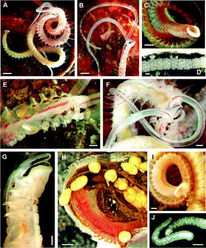
Fig. 2 (A) Polydora haswelli dorsal anterior SEM, palps regenerating, obscured prostomial tip bilobed as in Polydora websteri. (B) Polydora websteri dorsal anterior SEM. Arrows in A–B mark chaetiger 3 posterior boundaries. (C–D) Chaetiger 5 spines under light microscopy: (C) P. haswelli left side ventral view; (D) P. websteri in right side dorsal view. (E–F) SEM detail of pygidial fan inner surfaces: (E) P. haswelli fan; (F) P. websteri fan. Scale bars: A–B, 100 µm; C–D, 10 µm; E–F, 20 µm.
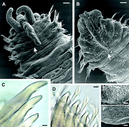
Fig. 3 Polydora haswelli chaetiger 5 spine SEM: (A) left side lateral view; (B) right side ventro-lateral view. Scale bars: 10 µm.
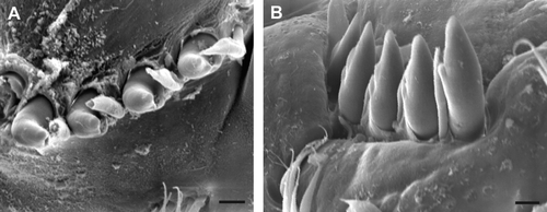
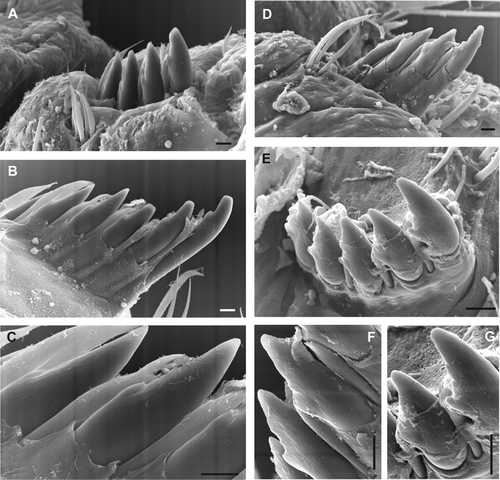
Fig. 4 Chaetiger 5 spine SEM, right side ventral views. (A–C) Polydora haswelli (2 specimens, B–C same specimen). (D–G) Polydora websteri (2 specimens, D & F, E & G same specimens). Chipping and grinding wear of the subdistal shelf is more pronounced for the wider shelf of the P. websteri spines where the lower contact edge has become irregular (image F & G). Scale bars: 10 µm.