340
Views
4
CrossRef citations to date
0
Altmetric
Research articles

Molecular characterisation of the complete nuclear ribosomal DNA sequence of the blacklip abalone Haliotis rubra

, , , &
Pages 430-443 | Received 30 Sep 2017, Accepted 18 Jan 2018, Published online: 05 Feb 2018

Figures & data

Figure 1. Eukaryotic ribosomal DNA structure.

Figure 1. Eukaryotic ribosomal DNA structure.

Table 1. Primers and polymerase chain reaction protocols used in this study.

Table 2. Collected information and GenBank accession numbers for abalone species used in this study.

Table 3. Nucleotide composition, length, and GenBank accession number of each rRNA region from H. rubra.

Table 4. Pairwise distances of 18S sequences (lower left matrix) and 28S rDNA sequences (upper right matrix).

Figure 2. A ML phylogenetic tree was constructed using the ‘GIR + G’ model with 1000 bootstrap replications. The numbers represent the bootstrap value (values <60% are not shown). 2n indicates the diploid chromosome number.
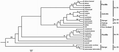
Figure 2. A ML phylogenetic tree was constructed using the ‘GIR + G’ model with 1000 bootstrap replications. The numbers represent the bootstrap value (values <60% are not shown). 2n indicates the diploid chromosome number.

Figure 3. The entire nucleotide sequence of the H. rubra rDNA IGS region. The repeated sequences are underlined as follows: single line for repeat type A and double line for repeat type B. A putative termination signal (poly(T) tract) is in the solid box, and the RNA polymerase I transcription promoter is in the dashed box.
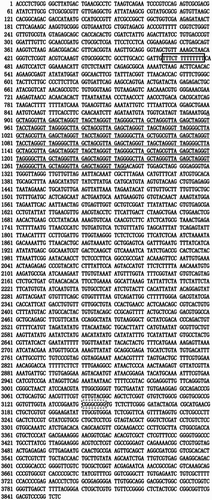
Figure 3. The entire nucleotide sequence of the H. rubra rDNA IGS region. The repeated sequences are underlined as follows: single line for repeat type A and double line for repeat type B. A putative termination signal (poly(T) tract) is in the solid box, and the RNA polymerase I transcription promoter is in the dashed box.

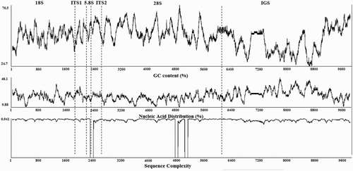
Figure 4. The GC content (%), nucleic acid distribution (% T), and sequence complexity determined using 80-bp windows across the entire rDNA nucleotide sequence of H. rubra.

Figure 4. The GC content (%), nucleic acid distribution (% T), and sequence complexity determined using 80-bp windows across the entire rDNA nucleotide sequence of H. rubra.

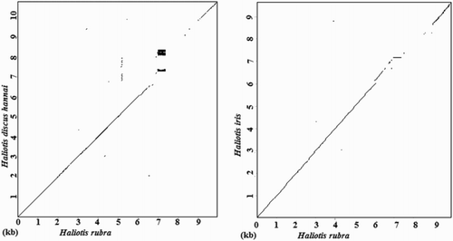
Figure 5. Dot-matrix comparisons of rDNA sequences. Dot plots indicate comparisons between H. rubra and H. discus hannai and between H. rubra and H. iris.

Figure 5. Dot-matrix comparisons of rDNA sequences. Dot plots indicate comparisons between H. rubra and H. discus hannai and between H. rubra and H. iris.

Reprints and Corporate Permissions

Please note: Selecting permissions does not provide access to the full text of the article, please see our help page How do I view content?

To request a reprint or corporate permissions for this article, please click on the relevant link below:

Academic Permissions

Please note: Selecting permissions does not provide access to the full text of the article, please see our help page How do I view content?

Obtain permissions instantly via Rightslink by clicking on the button below:

If you are unable to obtain permissions via Rightslink, please complete and submit this Permissions form. For more information, please visit our Permissions help page.