Figures & data
Table 1. Prevalence of abnormality phenotypes by region in 101 free-living New Zealand Chinook salmon.
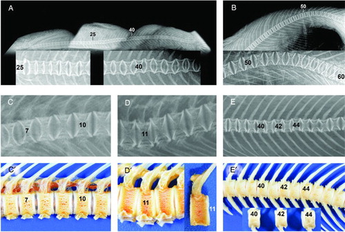
Figure 1. Examples of deformities found in free-living female Chinook salmon captured during spawning migrations up the Rangitata (A,B) and Waimakriri Rivers (C,D,E), in Canterbury, New Zealand. Types refer to the classification system of Witten et al. (Citation2009). A, A salmon with multiple deformities. Compressions of four vertebrae, numbers 26–29, and reduction of intervertebral spaces. Some loss of internal radiologic structure (left inset, Types 3,4). Vertical shifts (Type 17) of vertebrae numbers 39–43 (right inset). B, Kyphosis (Type 15) in the ural region without radiological changes in vertebral morphology. More caudal vertebrae normally show reduced length towards the tail fin. C and C’, Radiograph (C) of part of postcranial and abdominal regions showing dorsal vertical shifts (Type 17) of vertebrae 7–10 and in bone preparations (C’), showing uneven spacing between vertebrae. D and D’, Radiograph (D) and bone preparations (D’) showing lordosis (Type 14) at the level of vertebrae 10–12 and dorsal compression of vertebra 11. Vertebra 11 was 15.1% shorter dorsally than ventrally. E and E’, Radiograph (E) and bone preparations (E’) of multiple vertical shifts (Type 17) of vertebrae 40–46. Alternating compression dorsally and ventrally (see vertebrae 40,42,44) is more easily identified in articulated bones than in radiographs. Dorsal lengths of vertebrae 40,42,44 were significantly longer by 4.8% than ventral lengths (paired t-test, P = .017, d.f. = 2).