Figures & data
Figure 1. Aerial view of the mouth of the Waikouaiti River, Karitāne, north of Dunedin (−45.641023, 170.658784) showing drift accumulations of Bonnemaisonia estuarine shores with map inset of New Zealand.
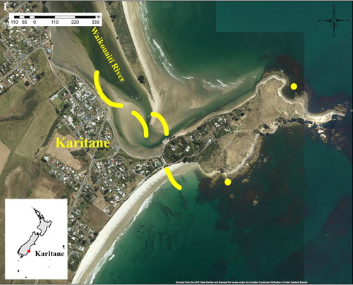
Figure 2. A, Aerial image of the mouth of the Waikouaiti River. B, Drift windrows of Bonnemaisonia – March 2019. C–D, Dense accumulations of Bonnemaisonia in shallow water in estuary – March 2019. E, Bonnemaisonia bloom showing growth as balls/pompoms –1 April 2019.
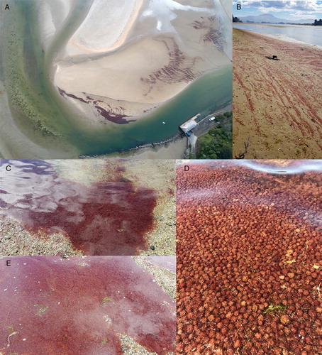
Figure 3. Subtidal habitats at Te Awa Mokihi (Butterfly Bay) in June 2019 with Bonnemaisonia hamifera. A, Numerous clumps settled on bottom associated with various turf and larger foliose algae. B, View of water column with numerous clumps of B. hamifera. C, Free-floating clumps of B. hamifera among Macrocystis forest. D, Non-native thallus of Undaria with B. hamifera.
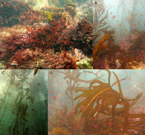
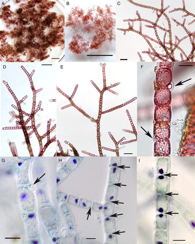
Figure 4. Field-collected samples of Bonnemaisonia hamifera. A-F, freshly collected; G-I, dead thalli. A–B, Clumps of B. hamifera showing irregular outlines. Scale bar = 1 cm. C–E, Filaments of B. hamifera with irregular to alternate branching. Scale bar = 100 µm. F–G, Portion of filaments with gland cells (arrows). Scale bar = 20 µm. H–I, Portions of filaments stained showing aniline blue with prominent, single rhodophysin granules in each cell, and pit connections between adjoining cells (arrows). Scale bar = 20 µm.