Figures & data
Table 1. Accession numbers for newly determined rDNA sequences of Bindiferia boggaya, B. fragilissima and Togula jolla collected at different sites
Figs 1–13. Light micrographs of Bindiferia fragilissima sp. nov. from France. Scale bars = 10 µm.
Figs 1, 2. Same actively motile cell in ventral view in different focal planes showing the sulcal path and cingulum; note the red stigma close to the apex (arrow) and the posterior nucleus (n).
Fig. 3. Actively motile cell in ventral view showing the sulcal path and cingulum; note the red stigma close to the apex (arrow).
Figs 4, 5. Same actively motile cell in ventral view showing the sulcal path including the extension onto the epicone (short arrow); note the red stigma close to the apex (arrow) and the length of the longitudinal flagellum.
Fig. 6. ‘Stationary motile’ cell; note the red stigma (arrow).
Fig. 7. ‘Stationary motile’ cell with two dorsal longitudinal grooves on the hypocone (arrowheads).
Figs 8, 9. Two ‘stationary motile’ cells with three dorsal longitudinal grooves on their hypocone (arrowheads); note the red stigma (arrow).
Fig. 10. Non-motile cell in ventral view covered by a hyaline layer (double arrowhead); note the red stigma (arrow).
Fig. 11. Non-motile division stage covered by a hyaline double-layer (double arrowhead); note the red stigma (arrow).
Fig. 12. Non-motile division stage with daughter cells starting to separate posteriorly, covered by a hyaline double-layer (double arrowhead); note the red stigma (arrow).
Fig. 13. Already motile, nearly divided, but still apically connected daughter cells outside the division cyst
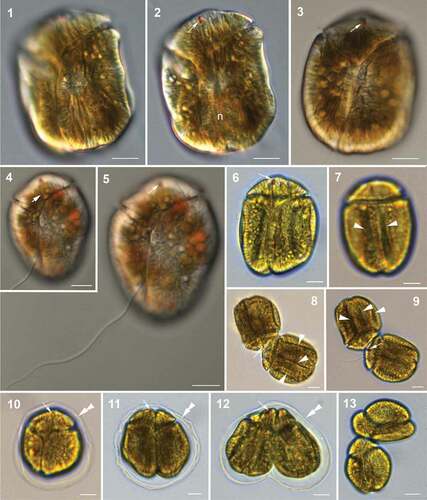
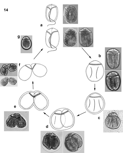
Fig. 14. Hypothetical vegetative life cycle (cell division) of Bindiferia fragilissima sp. nov. Free-swimming cells (a) have a smooth cell surface (lower illustrations) and sometimes many extrusomes can be visible along the cell periphery (upper illustrations). Stationary motile cells develop dorsal grooves (b) and get enclosed by a hyaline sheath (c). Vegetative binary fission takes place in the division cyst, with a hyaline sheath (d, e) and motile nearly divided but still apically connected daughter cells without surface grooves get released from the sheath (f). Smaller free-swimming cells with smooth surface (g) start growing
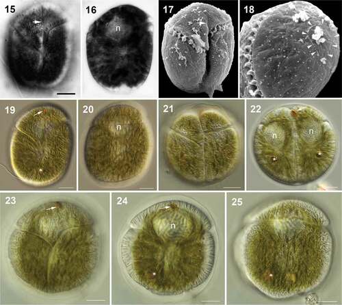
Figs 15–25. Light (15, 16, 19–25) and scanning electron (17, 18) micrographs of Bindiferia boggaya comb. nov. from Australia. Scale bars = 10 µm.
Figs 15–18. Cells from Sydney, Australia, from the original description (Murray & Patterson Citation2002).
Fig. 15. Cell in ventral view showing the cingulum and sulcus paths, note the sulcal extension (arrow).
Fig. 16. Cell in mid-focus showing the anteriorly located nucleus (n).
Fig. 17. Cell in ventral view showing the cingulum and sulcus paths; note the sulcal extension (arrow). The small arrowheads indicate the beginning and end points of the apical structure complex.
Fig. 18. Apical view of the epicone showing the loop-shaped apical structure complex (arrowheads).
Figs 19–25. Cells from Broome, Australia.
Fig. 19. Motile cell in ventral view; note the red stigma close to the apex (arrow) and possibly a food body (asterisk).
Fig. 20. Motile cell in dorsal view; note the anterior nucleus (n).
Figs 21, 22. Vegetative division stage in a hyaline sheath in different focal planes; note the red stigma (arrow), the nuclei (n) and possible food bodies (asterisk).
Figs 23–25. Non-motile cell in a hyaline sheath in different focal planes (23, ventral; 24 mid-focus; 25, dorsal); note the red stigma (arrow), the anterior nucleus (n) and a possible food body in the posterior area of the hypocone (asterisk). The dorsal hypocone surface is smooth, without grooves
Figs 26–31. Division stages within a hyaline sheath of Bindiferia boggaya comb. nov. from Broome, Australia (26–28) and New Zealand (29–31). Scale bars = 10 µm.
Figs 26, 29. Non-motile, rounded cell; note the stigma (arrow).
Figs 27, 30. Two daughter cells in the division cyst; note a stigma (arrow).
Figs 28, 31. Four daughter cells in the division cyst
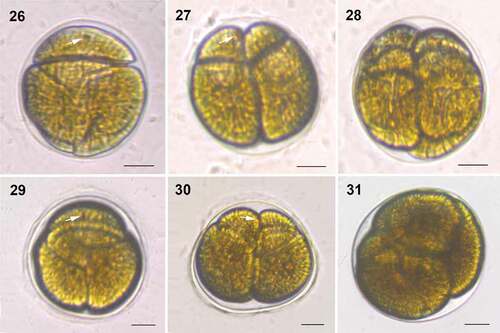
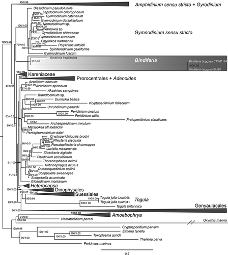
Fig. 32. Molecular phylogeny of the dinoflagellates, including the new sequences of Bindiferia and Togula, based on a concatenated alignment of 18S, ITS1, 5.8S, ITS2 and 28S rRNA gene regions. Most likely tree shown, with support values based on maximum likelihood bootstrap analysis (ML BS) and Bayesian inference posterior probabilities (BI PP)
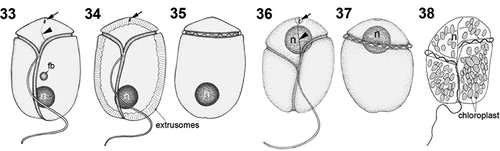
Figs 33–38. Line drawings of Bindiferia fragilissima (33–35) and B. boggaya (36–38). fb, food body; n, nucleus; arrows indicate stigma; arrowheads indicate sulcal extension.
Figs 33, 34, 36, 38. Ventral views.
Figs 35, 37. Dorsal views.
Fig. 38. Drawing from the original description (Murray & Patterson Citation2002)